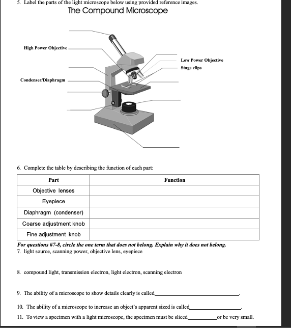
39 label the image of a compound light microscope using the terms provided
microscope - The compound microscope | Britannica The compound microscope. The limitations on resolution (and therefore magnifying power) imposed by the constraints of a simple microscope can be overcome by the use of a compound microscope, in which the image is relayed by two lens arrays. One of them, the objective, has a short focal length and is placed close to the object being examined.It is used to form a real image in the front focal ... PDF LAB 3 Use of the Microscope - Los Angeles Mission College LAB 3 - Use of the Microscope Introduction In this laboratory you will be learning how to use one of the most important tools in biology - the compound light microscope - to view a variety of specimens. You will also use a slightly different type of light microscope called a stereoscopic dissecting microscope.
A Study of the Microscope and its Functions With a Labeled Diagram The compound microscope uses light for illumination. Some compound microscopes make use of natural light, whereas others have an illuminator attached to the base. The specimen is placed on the stage and observed through different lenses of the microscope, which have varying magnification powers. Compound Microscope Parts and Functions

Label the image of a compound light microscope using the terms provided
Solved Microscope parts/labeling 9 Label the image of a - Chegg microscope parts/labeling 9 label the image of a compound light microscope using the terms provided. 1 points eyepiece eyepiece light source references references base arm slide holder arm stage mechanical stage fine adjustment knob power switch objectives mindeneer microscopy lab homework saved help save & exit submit eyepiece light source 9 … Solved Label the image of a compound light microscope using - Chegg Step-by-step answer. Who are the experts? Experts are tested by Chegg as specialists in their subject area. We review their content and use your feedback to keep the quality high. Transcribed image text: Label the image of a compound light microscope using the terms provided. Label the microscope - Science Learning Hub All microscopes share features in common. In this interactive, you can label the different parts of a microscope. Use this with the Microscope parts activity to help students identify and label the main parts of a microscope and then describe their functions. Drag and drop the text labels onto the microscope diagram.
Label the image of a compound light microscope using the terms provided. Microscope Notes - Northern Arizona University Microscope Drawings. When drawing what you see under the microscope, follow the format shown below. It is important to include a figure label and a subject title above the image. The species name (and common name if there is one) and the magnification at which you were viewing the object should be written below the image. Match the names of the microscope parts in column a Label the image of a compound light microscope using the terms provided. If leaving an objective lens over the stage when storing the microscope, which objective lens should be placed over the stage? A. High power 40X B. Scanning 4X C. Oil immersion 100XD. Low power 10X B. Scanning 4X B. Scanning 4X Light Microscope- Definition, Principle, Types, Parts, Labeled Diagram ... The difference is simple light microscopes use a single lens for magnification while compound lenses use two or more lenses for magnifications. This means, that a series of lenses are placed in an order such that, one lens magnifies the image further than the initial lens. The modern types of Light Microscopes include: Bright field Light Microscope Parts of the Microscope Printables - ThoughtCo Most microscopes used in a classroom setting are compound microscopes. These usually consist of a light source and three to five lenses with a total magnification of 40x to 1000x. The following free printables can help you teach your students the basic parts of a microscope so that they are ready to dive into a world previously unseen.
Labeling the Parts of the Microscope Labeling the Parts of the Microscope This activity has been designed for use in homes and schools. Each microscope layout (both blank and the version with answers) are available as PDF downloads. You can view a more in-depth review of each part of the microscope here. Download the Label the Parts of the Microscope PDF printable version here. Label the image to review the components of a compou... - Biology - Kunduz Label the image to review the components of a compound light microscope. Nosepiece Arm k Mechanical stage ces Base Stage adjustment Ocular Fine focus Diaphragm Light source Coarse focus Objective lens Wames Redfeam McGraw-Hill Education Reset < Previous Next > Show Answer Create an account. Get free access to expert answers A quick guide to light microscopy in cell biology - PMC Most cell biology imaging is done with widefield microscopy, in which the microscope simply forms an image of the sample on the camera, without any additional optical manipulation. Live cells are most commonly imaged on an inverted epifluorescence microscope (Figure 1). In such a microscope, the objective images the sample from below. Microscopy Lab Quiz Flashcards | Quizlet Label the image of a compound light microscope using the terms provided. If leaving an objective lens over the stage when storing the microscope, which objective lens should be placed over the stage? A. High power 40X B. Scanning 4X C. Oil immersion 100X D. Low power 10X B. Scanning 4X The fine adjustment knob on the microscope
Compound Microscope: Parts of Compound Microscope - BYJUS (A) Mechanical Parts of a Compound Microscope 1. Foot or base It is a U-shaped structure and supports the entire weight of the compound microscope. 2. Pillar It is a vertical projection. This stands by resting on the base and supports the stage. 3. Arm The entire microscope is handled by a strong and curved structure known as the arm. 4. Stage 2 using a compound light microscope observe each Using a compound light microscope, observe each smear. 4-1 Draw and label the appearance of each cell type (morphology and colony shape) in the space provided below. Indicate the total magnification next to each drawing. BIO 168 Module 2 Quiz Review Flashcards | Quizlet Each of the following steps are necessary in preparing and observing a wet mount. Place the steps in the correct order. 1. Obtain a clean slide and cover slip. 2. Using a transfer pipette, obtain a drop of specimen and place onto the center of the slide. PDF MICROSCOPE LAB - Yavapai College microscope. Types of microscopes: Light Microscope - the models found in most schools, use compound lenses and light to magnify objects. The lenses bend or refract the light, which makes the object beneath them appear closer. Scanning Electron Microscope - allow scientists to view a universe too small to be seen with a light microscope.
What is a Compound Microscope? - Microscope Manufacturer & Supplier A compound light microscope comes with its own source of light. The function of a light microscope is to target a beam of light on a specimen, to produce an image. The specimen you are studying must be thin and transparent. The image is positioned to pass through one or two lenses for greater magnification so that you get an enlarged view.
Working Principle and Parts of a Compound Microscope (with Diagrams) Therefore, the smallest details that can be seen by a typical light microscope is having the dimension of approximately 0.2 µ. Smaller objects or finer details than this cannot be resolved in a compound microscope. 5. Eyepiece: The eyepiece is a drum, which fits loosely into the draw tube.
Compound Light Microscopes Teaching Resources | Teachers Pay Teachers The Compound Light Microscope is a great introductory lesson on the basics of the microscope and how it is used. This product includes a labeled color poster of the microscope and how to use it as well as worksheets that include reading comprehension, labeling, fill in the blank and a sequence card set.
Parts of a microscope with functions and labeled diagram Q. List down the 18 parts of a Microscope. 1. Ocular Lens (Eye Piece) 2. Diopter Adjustment 3. Head 4. Nose Piece 5. Objective Lens 6. Arm (Carrying Handle) 7. Mechanical Stage 8. Stage Clip 9. Aperture 10. Diaphragm 11. Condenser 12. Coarse Adjustment 13. Fine Adjustment 14. Illuminator (Light Source) 15. Stage Controls 16. Base 17.
Label The Photomicrograph Using The Hints Provided / The Fate And ... Label the image of a compound light microscope using the terms provided. If temperature is increased, the rate of diffusion increases. Correctly label the following anatomical parts of a kidney. Potassium permanganate (molecular weight = 158) the difference in an area with high concentration and an area with low concentration is …
11 . O 2. Label the parts of the compound light micr... - Biology Names of these parts and. 11 . O 2. Label the parts of the compound light microscope on the diagram provided below (Figure 2-3). Names of these parts and their functions must be known to use the microscope correctly. ocular lens: remagnifies the image formed by the objective lens body tube: holds the lens system of the instrument arm: connects ...
Microscope Parts and Functions First, the purpose of a microscope is to magnify a small object or to magnify the fine details of a larger object in order to examine minute specimens that cannot be seen by the naked eye. Here are the important compound microscope parts... Eyepiece: The lens the viewer looks through to see the specimen.


Post a Comment for "39 label the image of a compound light microscope using the terms provided"