39 label the structures of the bone.
Bone Structure - CliffsNotes Compact bone consists of cylindrical units called osteons. Each osteon contains concentric lamellae (layers) of hard, calcified matrix with osteocytes (bone cells) lodged in lacunae (spaces) between the lamellae. Smaller canals, or canaliculi, radiate outward from a central canal, which contains blood vessels and nerve fibers. Bones: Types, structure, and function - Medical News Today Cancellous (trabecular or spongy) bone: This consists of a network of trabeculae or rod-like structures. It is lighter, less dense, and more flexible than compact bone. It is lighter, less dense ...
Solved Label the structures of the bone. Lesser trochanter - Chegg Question: Label the structures of the bone. Lesser trochanter Head Femur Popliteal surface Lateral epicondyle Intertrochanteric crest Neck Medial epicondyle Greater trochanter Reset Zoom This problem has been solved! See the answer fill in the blank Show transcribed image text Expert Answer 100% (12 ratings) Femur. It's none of lower limb.

Label the structures of the bone.
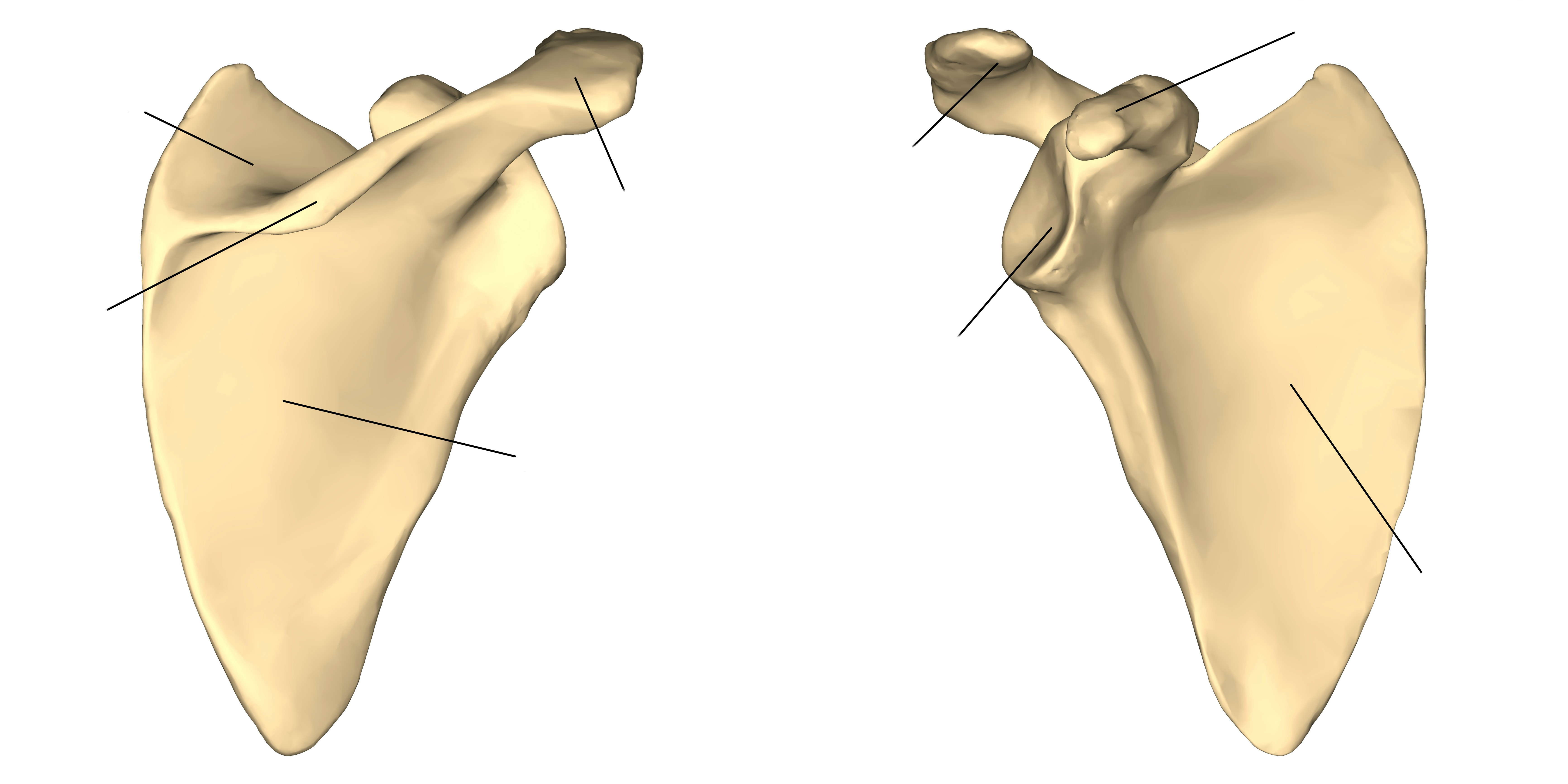
Anatomy & Physiology: Bone Labeling Flashcards | Quizlet Metatarsals Distal Phalanx Proximal Phalanx Calcaneous Navicular Cuboid Medial Cuneiform Intermediate Cuneiform Talus Lateral Cuneiform Scapula Acromion Supraspinous Fossa Spine Infraspinous Fossa Glenoid Fossa Caracoid Process Suprascapular Notch Inferior Angle Superior Angle Pelvic Gurdle Acetabulum Ilium Iliac Crest Greater Sciatic Notch Ischium Femur Bone Anatomy: Labeled Diagram and Quiz - EZmed Femur bone anatomy made easy using a labeled diagram of the main parts of the thigh bone along with their location. Includes anatomy of the femur quiz. ... The easy way to remember the name condyle is to imagine wrapping your hand around the condylar structure. Your hand will form a "C-shape". The "C-shape" will help you remember the ... A List of Bones in the Human Body With Labeled Diagrams It is one of the seven bones that form the orbital cavity, and consists of three parts. Facial Bones at a Glance Mandible (1) Maxilla (2) Palatine bone (2) Zygomatic bone (2) Nasal bone (2) Lacrimal bone (2) Inferior nasal conchae (2) Vomer (1) Total number of bones=14 Mandible This is the lower jawbone, and is known as the inferior maxillary bone.
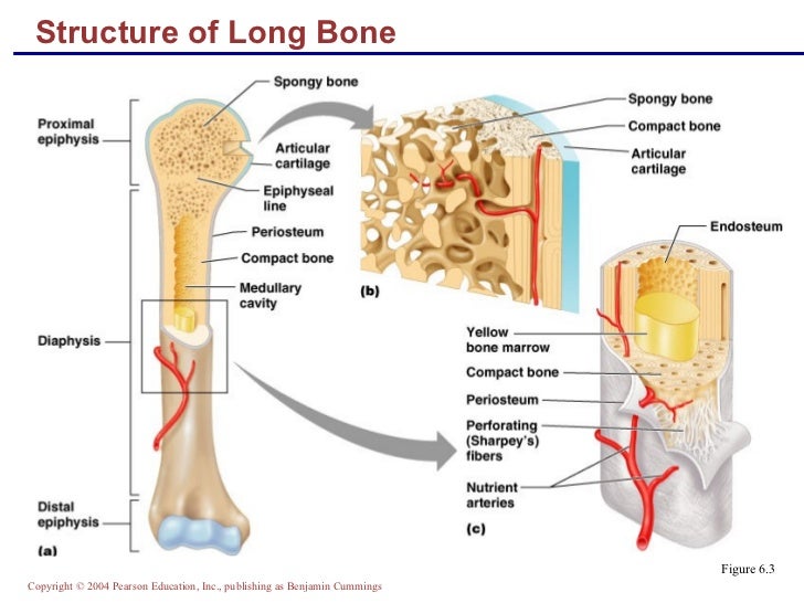
Label the structures of the bone.. Structure of the skeletal system - Skeletal system - OCR - GCSE ... The skeleton is the central structure of the body and is made up of bones, joints and cartilage. The skeleton provides the framework for muscles and gives the body its defined human shape. Part of Structure of the Long Bone Quiz - PurposeGames.com Labeling the structures of the long bone. Includes bone types and marrows. Remaining 0. Correct 0. Wrong 0. Press play! 0%. 0:00.0. Quit. Again. This game is part of a tournament. You need to be a group member to play the tournament. Join group, and play Just play. Your Scorecard. The scorecard of a champion. Score . 0 % Bone Structure | Anatomy and Physiology I | | Course Hero The diaphysis is the tubular shaft that runs between the proximal and distal ends of the bone. The hollow region in the diaphysis is called the medullary cavity, which is filled with yellow marrow. The walls of the diaphysis are composed of dense and hard compact bone. Figure 6.7. Anatomy of a Long Bone Solved Label the structures of the bones. Interosseous - Chegg Anatomy and Physiology questions and answers. Label the structures of the bones. Interosseous membrane Head of radius Radius Ulna Neck of radius Trochlear notch. Question: Label the structures of the bones.
Long bone anatomy, structure, parts, function and fracture types All of the bones in the arms and legs, except the patella, and bones of the wrist, and ankle, are long bones. Long bone structure A typical long bone consists of the following parts: The diaphysis (growing between) is the shaft of a long bone — the long, cylindrical, main portion of the bone. Label the following structures on the accompanying diagram of a long ... Label the following structures on the accompanying diagram of a long bone. diaphysis articular surfaces of the proximal end of the bone epiphysis articular surfaces of the distal end of the bone Expert's Answer Solution.pdf Next Previous Bone Structure & Anatomy Explained - What Is Bone Marrow? The structure of a long bone consists of several sections:. Diaphysis: This is the long central shaft. Epiphysis: Forms the larger rounded ends of long bones. Metaphysis: Area between the diaphysis and epiphysis at both ends of the bone. Epiphyseal Plates: Plates of cartilage, also known as growth plates which allow the long bones to grow during childhood. . Once we stop growing (between 18 ... Bone: Histology, constituents and types | Kenhub Bone is a modified form of connective tissue which is made of extracellular matrix, cells and fibers. The high concentration of calcium and phosphate based minerals throughout the connective tissue is responsible for its hard calcified nature.
Structure of a Long Bone - Level 2 anatomy and physiology The Long Bone is the most common type of bone in the human body The Structure of a Long Bone: Labelled Image Long bones are the most common bones found in the human body. They are composed mostly of compact bone, and are roughly cylindrical in shape with enlarged ends filled with spongy bone. Free Anatomy Quiz - The Skeletal System Section Bone anatomy - the structure of a bone : Quiz 1--- Quiz 2--- Quiz 3 Quiz 4-- Quiz 5. Resources : In this section we've added a few alternative study aids to help you along. Articles - Here you'll find a range of short articles on basic anatomy and physiology topics, complete with a few 'test yourself' questions for each one. 6.3 Bone Structure - Anatomy and Physiology 2e | OpenStax The outer surface of the bone is covered with a fibrous membrane called the periosteum (peri - = "around" or "surrounding"). The periosteum contains blood vessels, nerves, and lymphatic vessels that nourish compact bone. Tendons and ligaments also attach to bones at the periosteum. Lab 3 Flashcards | Quizlet Lab 3. -includes the skeletal elements within the limbs, as well as supporting the pectoral girdle and pelvic girdle. The shoulder girdle or pectoral girdle is the set of bones in the appendicular skeleton which connects to the arm on each side. In humans, it consists of the clavicle and scapula. Nice work!

Normal Anatomy of the Human Vertebral Column | Compel Visuals
6.3 Bone Structure - Anatomy & Physiology Bone Cells. Although bone cells compose less than 2% of the bone mass, they are crucial to the function of bones. Four types of cells are found within bone tissue: osteoblasts, osteocytes, osteogenic cells, and osteoclasts (Figure 6.3.5). Figure 6.3.5 - Bone Cells: Four types of cells are found within bone tissue. Osteogenic cells are undifferentiated and develop into osteoblasts.
Pre-Lab 2 – Human Anatomy Lab Manual
Structure of the skeletal system - Skeletal system - AQA - GCSE ... The skeleton is the central structure of the body and is made up of bones, joints and cartilage. The skeleton provides the framework for muscles and gives the body its defined human shape. Part of
ANAT2511 Basic Tissues - Embryology
Labeled Skeletal System Diagram - Bodytomy Bone Structure of the Hand There are around seven major bones from the shoulder to the palms. They are scapula, humerus, radius, ulna, carpels, metacarpals, and phalanges.

Bone Pictures II - Labeled Drawn | Chandler Physical Therapy
Skeletal System - Labeled Diagrams of the Human Skeleton The bones of the superior portion of the skull are known as the cranium and protect the brain from damage. The bones of the inferior and anterior portion of the skull are known as facial bones and support the eyes, nose, and mouth. Hyoid and Auditory Ossicles. The hyoid is a small, U-shaped bone found just inferior to the mandible. The hyoid is the only bone in the body that does not form a joint with any other bone—it is a floating bone.

Hints for BIO 122 Lab Quiz 3
Long Bone Anatomy: Structure and Parts of Long Bones Structure of an adult human long bone. The following image gets into a little more detail in regard to human long bone structure. The labels include periosteum, compact bone, nutrient artery & vein, medullary cavity, yellow bone marrow, endosteum, epiphyseal line, and spongy bone with red bone marrow. Structure of human bones explained

Chapter 6, Page 4 - HistologyOLM 4.0
Skeletal System: Parts, Structure, Functions, Bones, Videos, Examples The main function of the skeletal system is that it provides a framework to the body and provides shape. Along with the muscular system, the skeletal system helps in the movement of the body parts of the body and locomotion of the body. The skeletal system is hard and so forms a protective layer for the softer, more delicate organs from any ...
What does "bone structure" mean? | Socratic
Bone Structure - Anatomy and Physiology The outer surface of bone, except in regions covered with articular cartilage, is covered with a fibrous membrane called the periosteum. Flat bones consist of two layers of compact bone surrounding a layer of spongy bone. Bone markings depend on the function and location of bones. Articulations are places where two bones meet.
![Untitled Document [bio.sunyorange.edu]](http://bio.sunyorange.edu/updated2/THINKING_EVOLUTION/anatomy1a/skull/teeth_op2.jpg)
Untitled Document [bio.sunyorange.edu]
Structure of Bone Tissue | SEER Training There are two types of bone tissue: compact and spongy. The names imply that the two types differ in density, or how tightly the tissue is packed together. There are three types of cells that contribute to bone homeostasis. Osteoblasts are bone-forming cell, osteoclasts resorb or break down bone, and osteocytes are mature bone cells. An equilibrium between osteoblasts and osteoclasts maintains bone tissue.
Post a Comment for "39 label the structures of the bone."