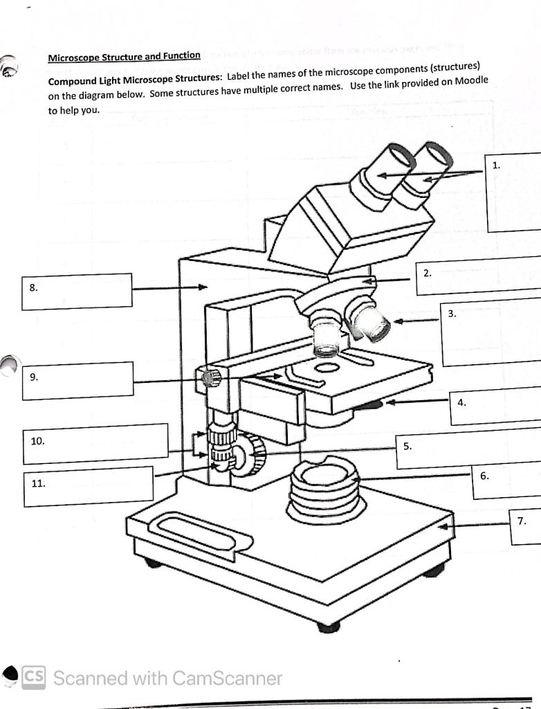
39 microscope drawing with label and function
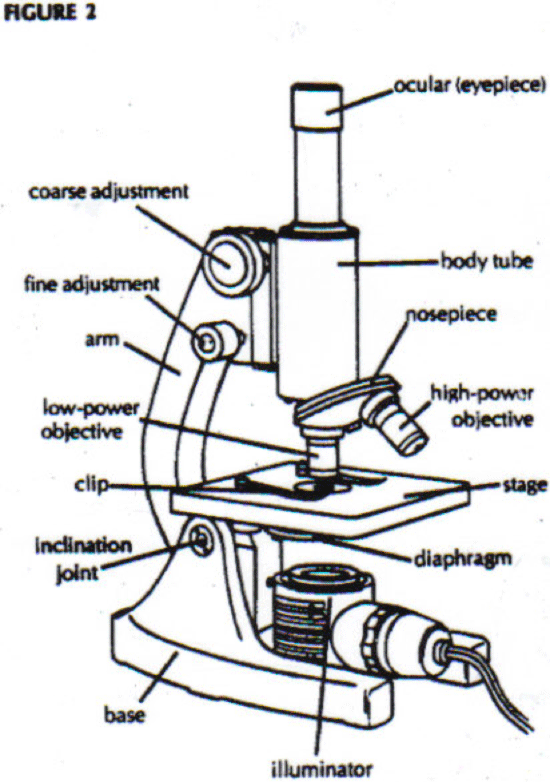
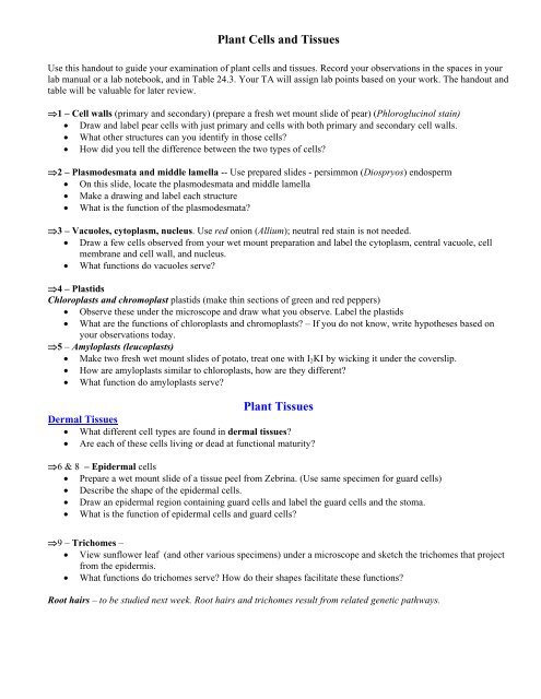
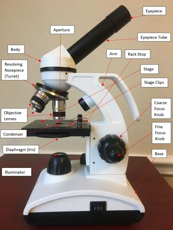
Label Microscope Diagram - EnchantedLearning.com arm - this attaches the eyepiece and body tube to the base. base - this supports the microscope. body tube - the tube that supports the eyepiece. coarse focus adjustment - a knob that makes large adjustments to the focus. diaphragm - an adjustable opening under the stage, allowing different amounts of light onto the stage. 16 Parts of a Compound Microscope: Diagrams and Video Once you have an understanding of the parts of the microscope it will be much easier to navigate around and begin observing your specimen, which is the fun part! The 16 core parts of a compound microscope are: Head (Body) Arm. Base. Eyepiece. Eyepiece tube.
Pin on Science worksheets - Pinterest A Study of the Microscope and its Functions With a Labeled Diagram To better understand the structure and function of a microscope, we need to take a look at the labeled microscope diagrams of the compound and electron microscope. These diagrams clearly explain the functioning of the microscopes along with their respective parts. M mooketsi

Microscope drawing with label and function
PDF Parts of a Microscope Printables - Homeschool Creations Label the parts of the microscope. You can use the word bank below to fill in the blanks or cut and paste the words at the bottom. Microscope Created by Jolanthe @ HomeschoolCreations.net. Parts of a eyepiece arm stageclips nosepiece focusing knobs illuminator stage objective lenses Label the microscope — Science Learning Hub Use this with the Microscope parts activity to help students identify and label the main parts of a microscope and then describe their functions. Drag and drop the text labels onto the microscope diagram. If you want to redo an answer, click on the box and the answer will go back to the top so you can move it to another box. Microscope drawing Images, Stock Photos & Vectors - Shutterstock Find Microscope drawing stock images in HD and millions of other royalty-free stock photos, illustrations and vectors in the Shutterstock collection. Thousands of new, high-quality pictures added every day.
Microscope drawing with label and function. Compound Microscope- Definition, Labeled Diagram, Principle, Parts, Uses The naked eye can now view the specimen at magnification 400 times greater and so microscopic details are revealed. Alternatively, the magnification of the compound microscope is given by: m = D/ fo * L/fe where, D = Least distance of distinct vision (25 cm) L = Length of the microscope tube fo = Focal length of the objective lens Solved Draw, label and write the functions of the parts of | Chegg.com How to use microscope before, during and after Turn the revolving turret so that the lowest power objective lens (eg. 4x) is clicked into position. Place the microscope slide on the stage and fasten it with the stage clips. Look at the objective lens… View the full answer Microscope Drawing: How to Sketch Microscope Slides Outline the general shapes: Draw the outline of largest shape onto the paper, making it fit within the quarters. Keep you pencil drawings light and adjust the shape as needed. This may require going between the microscope slide and the drawing in order to get the proportions and shape correct. Now move to the other shapes in your field of view. Compound Microscope Parts - Labeled Diagram and their Functions - Rs ... The eyepiece (or ocular lens) is the lens part at the top of a microscope that the viewer looks through. The standard eyepiece has a magnification of 10x. You may exchange with an optional eyepiece ranging from 5x - 30x. [In this figure] The structure inside an eyepiece. The current design of the eyepiece is no longer a single convex lens.
Parts Of The Microscope Label Worksheets & Teaching Resources | TpT Check out this well-organized Microscope label and describe worksheet. ... Help kids learn parts & functions using 3 different microscope diagrams, part labels & function description cards. ... so I chose to have my students focus on slides of various samples of bacteria cells. This provides the "cell drawing" outlines that students will need ... How to Sketch a Microscope Slide - Identifying and Sketching Cell ... Sketches come to life when you add highlights, shadows and color. For a pencil sketch, separate areas into white, light, medium and dark grey and black. To see the light/dark areas, squint so that the hard edges are blurred and your focus is on the shading. Start shading the light areas by following the shapes. Labeling the Parts of the Microscope Microscope World explains the parts of the microscope, including a printable worksheet for schools and home. Need Asssistance? 800-942-0528. Microscope Blog ... Labeling the Parts of the Microscope. This activity has been designed for use in homes and schools. Each microscope layout (both blank and the version with answers) are available as PDF ... Microscope Parts, Function, & Labeled Diagram - slidingmotion Microscope parts labeled diagram gives us all the information about its parts and their position in the microscope. Microscope Parts Labeled Diagram The principle of the Microscope gives you an exact reason to use it. It works on the 3 principles. Magnification Resolving Power Numerical Aperture. Parts of Microscope Head Base Arm Eyepiece Lens
Labeling the Parts of the Microscope | Microscope activity, Science ... Jan 13, 2016 - Free worksheets for labeling parts of the microscope including a worksheet that is blank and one with answers. How To Draw A Microscope - YouTube Today, we're learning how to draw a cool microscope!👩🎨 JOIN OUR ART HUB MEMBERSHIP! VISIT 🎨 VISIT OUR AMAZON ART SUPPLY S... Simple Microscope - Parts, Functions, Diagram and Labelling Simple Microscope - Parts, Functions, Diagram and Labelling A microscope is one of the commonly used equipment in a laboratory setting. A microscope is an optical instrument used to magnify an image of a tiny object; objects that are not visible to the human eyes. Table of Contents The common types of microscopes are: What is a Simple microscope? Compound Microscope: Definition, Diagram, Parts, Uses, Working ... - BYJUS The compound microscope is mainly used for studying the structural details of cell, tissue, or sections of organs. The parts of a compound microscope can be classified into two: Non-optical parts Optical parts Non-optical parts Base The base is also known as the foot which is either U or horseshoe-shaped.
Microscopes & Magnification | Optics Quiz - Quizizz
Microscope, Microscope Parts, Labeled Diagram, and Functions Microscope, Microscope Parts, Labeled Diagram, and Functions What is Microscope? A microscope is a laboratory instrument used to examine objects that are too small to be seen by the naked eye. It is derived from Ancient Greek words and composed of mikrós, "small" and skopeîn,"to look" or "see".
Alternate grade 11 - ccbbiology11
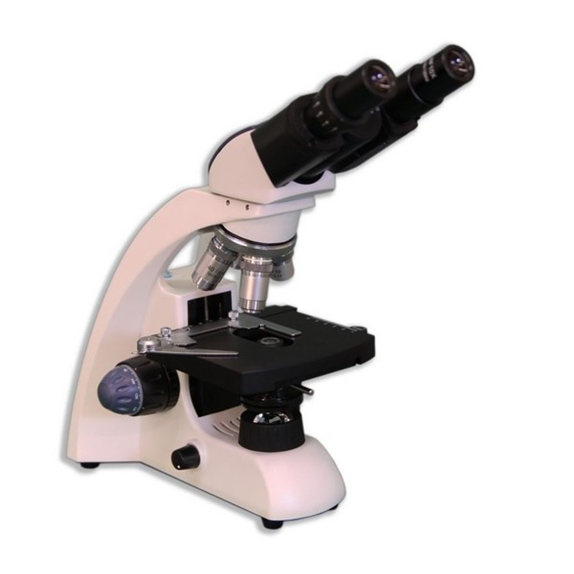
Microscope Parts & Functions - AmScope Microscope Parts and Functions Invented by a Dutch spectacle maker in the late 16th century, compound light microscopes use two sets of lenses to magnify images for study and observation. The first set of lenses are the oculars, or eyepieces, that the viewer looks into; the second set of lenses are the objectives, which are closest to the specimen.

Microscope, Microscope Parts, Labeled Diagram, and Functions
Microscope Types (with labeled diagrams) and Functions A compound microscope: Is used to view samples that are not visible to the naked eye. Uses two types of lenses - Objective and ocular lenses. Has a higher level of magnification - Typically up to 2000x. Is used in hospitals and forensic labs by scientists, biologists and researchers to study micro organisms. Compound microscope labeled diagram.
Microscope Gifts & Merchandise | Redbubble
A Study of the Microscope and its Functions With a Labeled Diagram To better understand the structure and function of a microscope, we need to take a look at the labeled microscope diagrams of the compound and electron microscope. These diagrams clearly explain the functioning of the microscopes along with their respective parts. Man's curiosity has led to great inventions. The microscope is one of them.

Microscope Diagram And Functions - Free Clipart Images ...
Microscope Parts and Functions First, the purpose of a microscope is to magnify a small object or to magnify the fine details of a larger object in order to examine minute specimens that cannot be seen by the naked eye. Here are the important compound microscope parts... Eyepiece: The lens the viewer looks through to see the specimen.

Microscope Diagram (Structures & Functions) Diagram | Quizlet
How to draw compound of Microscope easily - step by step - YouTube I will show you " How to draw compound of microscope easily - step by step "Please watch carefully and try this okay.Thanks for watching.....#microscopedrawi...

Microscope Parts and Functions
Parts of a Compound Microscope and Their Functions Body Tube: It is the tubular structure of microscope, hollow component of the microscope arm that is attached to the top half of the arm.With the use of adjustment knobs, it can be adjusted up and down. Nose Piece: It's a spinning metal element affixed to the lower end of the body tube in a circular pattern.It has three holes for objective lenses to be inserted into.
Microscope Parts & Function - ppt video online download
Parts of a microscope with functions and labeled diagram Optical parts of a microscope and their functions The optical parts of the microscope are used to view, magnify, and produce an image from a specimen placed on a slide. These parts include: Eyepiece - also known as the ocular. This is the part used to look through the microscope. Its found at the top of the microscope.
Free Microscope Drawing, Download Free Microscope Drawing png ...
Parts of the Microscope with Labeling (also Free Printouts) Let us take a look at the different parts of microscopes and their respective functions. 1. Eyepiece it is the topmost part of the microscope. Through the eyepiece, you can visualize the object being studied. Its magnification capacity ranges between 10 and 15 times. 2. Body tube/Head It is the structure that connects the eyepiece to the lenses.

Compound Microscope Parts – Labeled Diagram and their ...
Compound Microscope Parts, Functions, and Labeled Diagram Compound Microscope Parts, Functions, and Labeled Diagram Parts of a Compound Microscope Each part of the compound microscope serves its own unique function, with each being important to the function of the scope as a whole.

Compound Microscope: Parts of Compound Microscope
Labelled Diagram of Compound Microscope - Biology Discussion The below mentioned article provides a labelled diagram of compound microscope. Part # 1. The Stand: The stand is made up of a heavy foot which carries a curved inclinable limb or arm bearing the body tube. The foot is generally horse shoe-shaped structure (Fig. 2) which rests on table top or any other surface on which the microscope in kept.

BIOLOGY FROM 1 | EQUIPMENTS USED FOR OBSERVATION | Cours ...
Microscope drawing Images, Stock Photos & Vectors - Shutterstock Find Microscope drawing stock images in HD and millions of other royalty-free stock photos, illustrations and vectors in the Shutterstock collection. Thousands of new, high-quality pictures added every day.

22 Parts Of a Microscope With Their Function And Labeled ...
Label the microscope — Science Learning Hub Use this with the Microscope parts activity to help students identify and label the main parts of a microscope and then describe their functions. Drag and drop the text labels onto the microscope diagram. If you want to redo an answer, click on the box and the answer will go back to the top so you can move it to another box.

Compound Microscope Parts, Diagram Definition, Application ...
PDF Parts of a Microscope Printables - Homeschool Creations Label the parts of the microscope. You can use the word bank below to fill in the blanks or cut and paste the words at the bottom. Microscope Created by Jolanthe @ HomeschoolCreations.net. Parts of a eyepiece arm stageclips nosepiece focusing knobs illuminator stage objective lenses

A schematic drawing of the electrical cell in combination ...

Dissecting Stereo Microscope Parts and Functions
Microscope Diagram And Functions - Free Clipart Images ...

label microscope diagram | Charts | Microscope, Anatomy bones ...

Motic BA310 Digital Biological Microscope
Compound Microscope Parts, Functions, and Labeled Diagram ...
Microscope Parts and Functions
Solved 2. Calculate total magnification and measure field of ...

Parts of a Microscope Labeling Activity

Getting to Know the Microscope | Manualzz

Label the microscope — Science Learning Hub

Compound Microscope Parts

Parts of Stereo Microscope (Dissecting microscope) – labeled ...
Plant Cells and Tissues

The Cell: Parts and Functions for Kids – HowForKids

How to draw Compound microscope | Microscope diagram easily Draw | SSC diagram

Compound Microscope Parts, Functions, and Labeled Diagram ...

Microscopy | Introducing the Cell

Simple Microscope - Diagram (Parts labelled), Principle ...
Lesson 2 - The Light Microscope - Year 8 ScienceClass
Parts of the Microscope with Labeling (also Free Printouts ...
16 Parts of a Compound Microscope: Diagrams and Video ...

Parts of a microscope with functions and labeled diagram
Light Microscope- Definition, Principle, Types, Parts ...

Microscope Diagram Labeled, Unlabeled and Blank | Parts of a ...
Post a Comment for "39 microscope drawing with label and function"