42 clam labeled diagram
PDF Clam Anatomy Exercise - Florida Shellfish Aquaculture Online Resource Guide Clam Anatomy Exercise . Objective: Students will observe the inside and outside of a clam. Record on the sheet provided. Each student will receive the following materials: fresh clam (in shell), shucked clam, calipers, tray, dissecting tools, clam diagrams . Outside of the clam shell (use live clam): Third grade Lesson This Invertebrate is Clamtastic! - BetterLesson During this lesson, students get up close and personal with a very common invertebrate, the clam.The purpose of this lesson is for my students to see and feel the characteristics of an invertebrate, while completing a diagram of the clam's shell as well as its anatomy. I try to infuse as much academic vocabulary as possible into our discussions ...
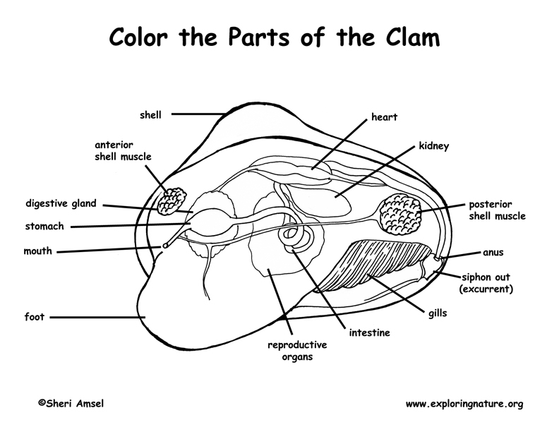
Clam Anatomy Labeling Page - Exploring Nature Clam Anatomy Labeling Page. Higher Resolution PDF for Printing. Click Here. Use Teacher Login to show answer keys or other teacher-only items. Link to More Info About this Animal (with Labeled Body Diagram) Click Here. Citing Research References. When you research information you must cite the reference. Citing for websites is different from ...

Clam labeled diagram
PDF Clam Dissection Guideline - Monadnock Regional High School Clam Dissection Guideline BACKGROUND: Clams are bivalves, meaning that they have shells consisting of two halves, or valves.The valves are joined at the top, and the adductor muscles on each side hold the shell closed. If the adductor muscles are relaxed, the shell is pulled open by ligaments located on each side of the umbo.The clam's foot is used to dig down into the Clam Diagram & Parts | What Is a Clam? | Study.com Belonging to a diverse group of animals known as bivalves, clams can be identified by the presence of two valves, or shells, joined by a hinge that allows the two shells to open or close. A... New Page 1 [ez002.k12.sd.us] Using the words in the above table label the following diagrams of the clam. C. Use arrows on the clam diagram to trace the pathway of food as it travels to the clam's stomach. Continue the arrows showing wastes leaving through the anus. Name the CLAM PHYLUM __MOLLUSK________________it means SOFT BODIED
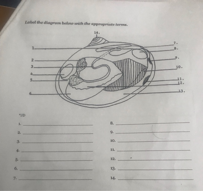
Clam labeled diagram. DOC Clam Dissection - PC\|MAC 25. Answer the questions on your lab report & label the diagrams of the internal structures of the clam. Also, use arrows on the clam diagram to trace the pathway of food as it travels to the clam's stomach. Continue the arrows showing wastes leaving through the anus. Clam Dissection Questions. Lab Questions: 1. Clam Anatomy Diagram | Quizlet large muscles that hold the shell together. heart. A hollow, muscular organ that pumps blood throughout the body. muscular foot. used for movement. mantle. secretes mother of pearl; surrounds and protects the soft body of molluscs. palp. moves food particles to mouth in molluscs. Bivalve (Clam) Diagram Quiz - PurposeGames.com About this Quiz. This is an online quiz called Bivalve (Clam) Diagram. There is a printable worksheet available for download here so you can take the quiz with pen and paper. This quiz has tags. Click on the tags below to find other quizzes on the same subject. biology. Clam Dissection - BIOLOGY JUNCTION Answer the questions on your lab report & label the diagrams of the internal structures of the clam. Also, use arrows on the clam diagram to trace the pathway of food as it travels to the clam's stomach. Continue the arrows showing wastes leaving through the anus. CLICK HERE FOR LAB QUESTIONS CLICK HERE FOR PHOTOS/QUIZ
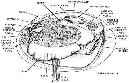
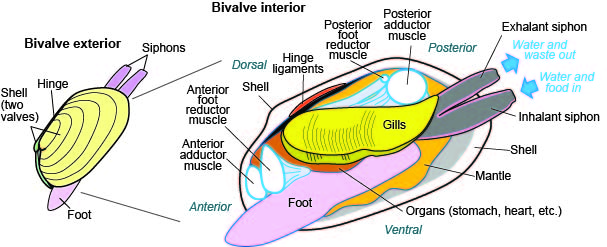
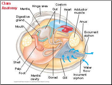
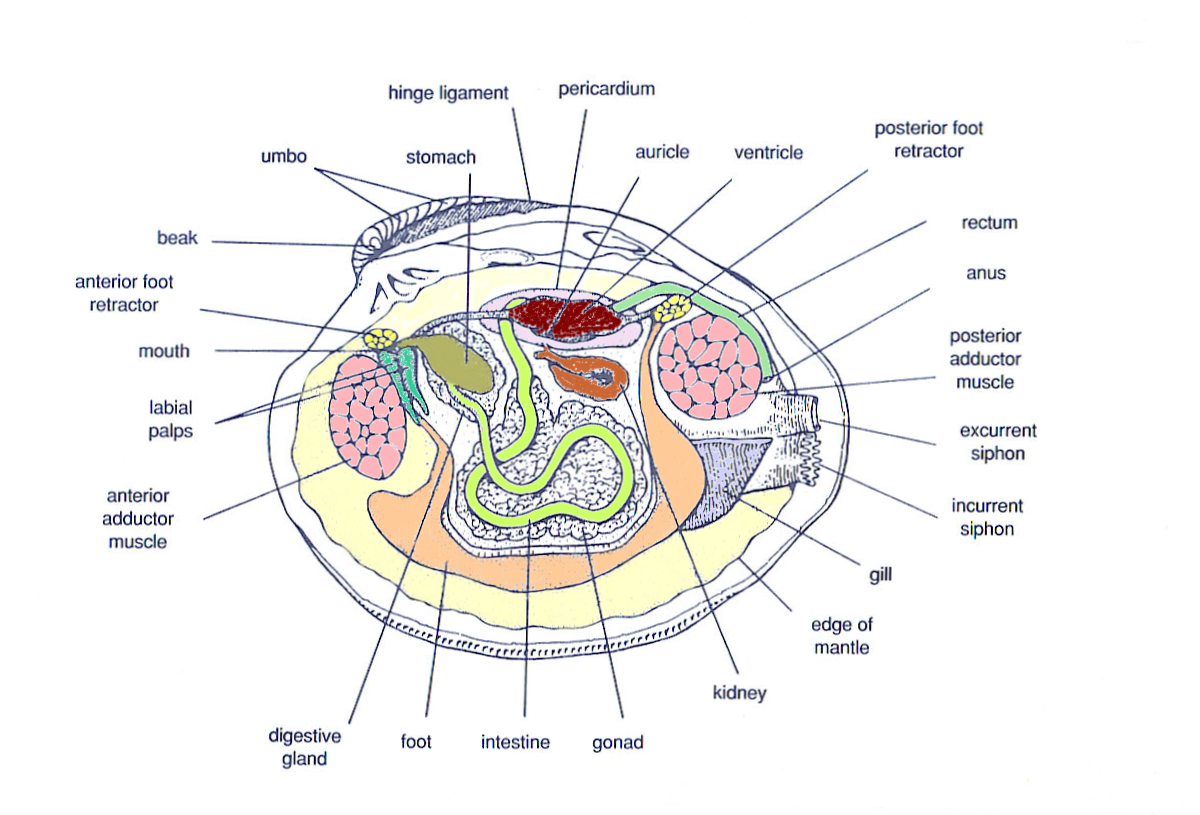
internal anatomy of a clam clam labeling anatomy exploringnature. Clam Dissection (or Mussel) | Ingridscience.ca . clam dissection mussel ingridscience. Images For BIO 122 Lab klemow.wilkes.edu. dissection labeled dissected ventral grasshopper lab nerve cord male dorsal female mouth showing unlabeled lateral wilkes edu views. Anatomy Of Animals ... PDF Biology of Bivalve Molluscs - Florida Shellfish Aquaculture Online ... Internal Clam Shell Anatomy 1. Mantle •Covers visceral or body mass •Holds in fluid •Secrets new shell 2. Ant. adductor muscle 3. Post. adductor muscle •Hold valves shut 4. Pericardium cavity •Region covered with thin, dark membrane •Contains 2-chambered heart and kidney in a fluid-filled sac 5. Mantle edge Clam Dissection Diagram | Quizlet digestive gland. green mush in visceral mass. gonad. pink mush in visceral mass. intestine. yellow mush in visceral mass. Latin Phrases. rjli012. Exam 3 - Blood supply of arteries. PDF Taxonomy, Anatomy, and Biology of the Hard Clam Internal Clam 1. Inner surface of left valve 2 Pt dd t l Shell Anatomy Post. adductor muscle 3. Ant. adductor muscle •Hold valves shut 4. Hinges •Ligament holds valves together •Interlocking teeth prevent valves from side slipping when opening and closing 5. Tth Teeth along ventral margin •Prevent valves from sliding when closes 6.
clam anatomy Diagram | Quizlet Start studying clam anatomy. Learn vocabulary, terms, and more with flashcards, games, and other study tools. Lab Bio: Clams Diagram | Quizlet a tooth of the hinge of a bivalve mollusk's shell situated just under the umbo and often relatively large. Gonad of Clam. A sex gland or reproductive gland. Mantle of Clam. protects the clam from sunlight in shelled mollusks, the the organ that forms the shell, and adds to the shell to increase its size and strength as the animal grows. anatomy of clam - Microsoft geoduck clam cooking prepare Index Of /images barnegatshellfish.org clam diagram labeled internal structures anatomy01 label incurrent excurrent siphon lab diagrams answer Images For BIO 122 Lab klemow.wilkes.edu unlabeled dissected lab earthworm labeled anterior wilkes dorsal bio Clam Anatomy (Function Quiz) clam Leevonk.com Clam Diagram Quiz - By dwhite298 - Sporcle 4. Element Symbols: True or False? II. 5. Badly Sculpted Animals - Invertebrates. 6. Square Numbers Scrambled (1-50) 7. Typing Challenge: Moons of our Solar System.
Phylum Mollusca | Geologic Overview of the Trenton Group
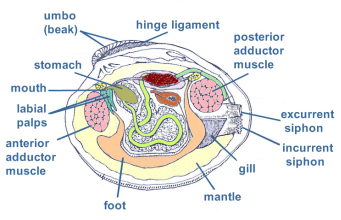
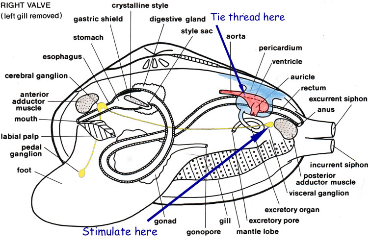
PDF Investigation #5 - Clam Anatomy - COSEE Locate the following parts of your clam according to the diagram: adductor muscles gills mantle excurrent siphon incurrent siphon stomach mouth foot intestine Lift the gills to find the stomach and intestines. Insert the skewer into the mouth and see that it empties into the stomach. Locate the foot that is used for digging.
Hard clams - Barnegat Bay
clam shell anatomy clam anatomy clams external flickr diagram foot body breakdown licking viral scientific salt courtesy fresh water. Pictures - Mollusks 101 sites.google.com. diagram clams mollusca bivalvia mussels phylum bivalve clam class gastropods bivalves scallops oysters mollusks cephalopods mussel parts higher label resolution.

Basic Clam Anatomy (Internal) Quiz
New Page 1 [ez002.k12.sd.us] Using the words in the above table label the following diagrams of the clam. C. Use arrows on the clam diagram to trace the pathway of food as it travels to the clam's stomach. Continue the arrows showing wastes leaving through the anus. Name the CLAM PHYLUM __MOLLUSK________________it means SOFT BODIED

Quia - Unit 4: Mollusks, Arthropods and Echinoderms
Clam Diagram & Parts | What Is a Clam? | Study.com Belonging to a diverse group of animals known as bivalves, clams can be identified by the presence of two valves, or shells, joined by a hinge that allows the two shells to open or close. A...

Clam Anatomy - Clam Discussion - Nano-Reef Community
PDF Clam Dissection Guideline - Monadnock Regional High School Clam Dissection Guideline BACKGROUND: Clams are bivalves, meaning that they have shells consisting of two halves, or valves.The valves are joined at the top, and the adductor muscles on each side hold the shell closed. If the adductor muscles are relaxed, the shell is pulled open by ligaments located on each side of the umbo.The clam's foot is used to dig down into the

Giant Clam (Tridacna gigas) - Alex, Ryan, and Sam; Muscular ...
Solved Class Bivalvia (clams, scallops, ovsters and mussels ...

clam Diagram | Quizlet

clam anatomy101 | Clam Anatomy Diagram | tanks4thememories ...
![Zoölogy [microform] : descriptive and practical. Zoology ...](https://c8.alamy.com/comp/REND9E/zology-microform-descriptive-and-practical-zoology-zoologie-pelecypodt-veo-cose-quoteatthr-t-accomplished-here-quotpraton-is-undoubtedly-the-structure-of-the-glllfl-t7ou-hi-0-a-in-sineaere-hr-t-artery-anterior-adduclt-tor-inuscie-auricle-ventricle-poiterior-adductor-mukl-fig-67-body-of-clam-left-valve-removed-double-walled-and-a-cross-section-is-like-a-letter-v-t-j-1-quot-quot-v-shaped-pocket-or-trough-the-to-quott-jquotquot-rquotj-p-quotquot-quoty-cross-partitions!-sectio7aw-t-REND9E.jpg)
Zoölogy [microform] : descriptive and practical. Zoology ...
MARINE BIOLOGY: CLAM DISSECTION

Clam Anatomy Panel – The Science Bank

The hatchery culture of bivalves: a practical manual
Untitled 1

bioweb images | Marine biology, Anatomy, Animal science

diagram of clam organs | Fossil Lady
Bivalves ~ New Jersey Scuba Diving

Mollusks Clam dissection & Bivalve information - ppt download
Untitled 1

Clam interior | Synergy Middle School Science 08-09

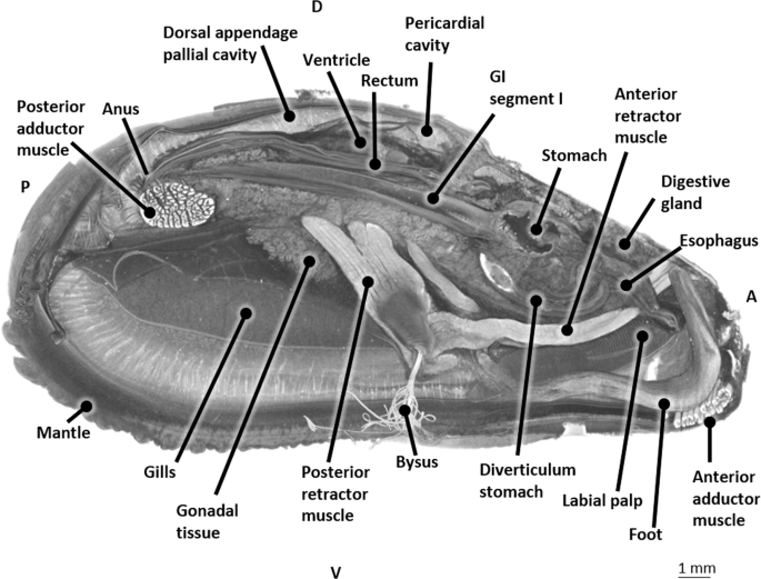
The internal, soft tissue anatomy of clam | Download ...

Clam Dissection - BIOLOGY JUNCTION
Solved] Draw a diagram and label the internal anatomy of the ...

Game Statistics - Anatomy of a Freshwater Clam
Clam Anatomy Coloring Page
Clam Dissection
Mussels and Clams (Bivalvia)
lab_append
Bivalves (pelecypods, clams, etc.), Fossils, Kentucky ...

Incredible Anatomy Of A Clam References - PeepsBurgh

Butter clam tissues for this study. A. Diagram of butter clam ...
Clam Dissection
The blue mussel inside: 3D visualization and description of ...
MARINE BIOLOGY: CLAM DISSECTION

Clam Anatomy Diagram | Quizlet
Clam Dissection - BIOLOGY JUNCTION

clam anatomy worksheet in 2022 | Color worksheets, Animal ...

Clam diagram Flashcards | Quizlet
Bivalves

New Page 1

Bivalve (Clam) Diagram Quiz
Hard clams - Barnegat Bay
Post a Comment for "42 clam labeled diagram"