45 labelled diagram of microscope
Label the Microscope Diagram | Download Scientific Diagram - ResearchGate Download scientific diagram | Label the Microscope Diagram from publication: Laboratory Exercises in Microbiology: Discovering the Unseen World through Hands-on Investigation | Microbiology ... Microscope Labeling - The Biology Corner Students label the parts of the microscope in this photo of a basic laboratory light microscope. Can be used for practice or as a quiz. ... 20. A microscope has an ocular objective of 10x and a high power objective of 50x, what is the microscope's total magnification? _____
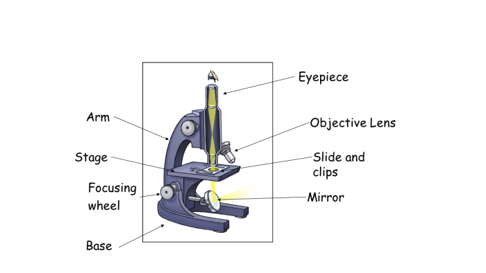
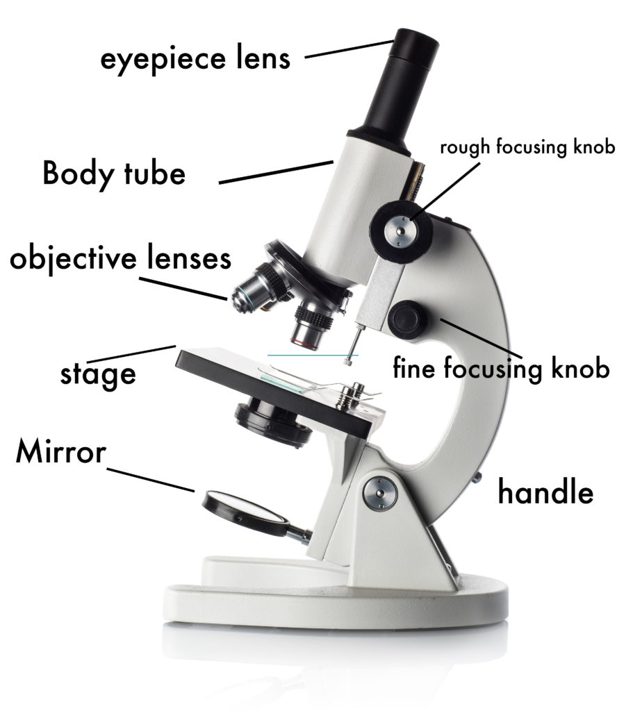
Label Microscope Diagram - EnchantedLearning.com base - this supports the microscope. body tube - the tube that supports the eyepiece. coarse focus adjustment - a knob that makes large adjustments to the focus. diaphragm - an adjustable opening under the stage, allowing different amounts of light onto the stage. eyepiece - where you place your eye.

Labelled diagram of microscope
Microscope Parts, Function, & Labeled Diagram - slidingmotion Microscope parts labeled diagram gives us all the information about its parts and their position in the microscope. Microscope Parts Labeled Diagram The principle of the Microscope gives you an exact reason to use it. It works on the 3 principles. Magnification Resolving Power Numerical Aperture. Parts of Microscope Head Base Arm Eyepiece Lens Investigating cells with a light microscope - BBC Bitesize Record the microscope images using labelled diagrams or produce digital images. When first examining cells or tissues with low power, draw an image at this stage, even if going on to examine the ... Microscope Types (with labeled diagrams) and Functions Phase-contrast microscope labeled diagram Phase-contrast microscope functions: Its applications areas include In cases where the specimen is colorless and is very tiny In biology to conduct cellular level examination of microorganisms that can't be visualized using the bright field microscopy Interference Microscope
Labelled diagram of microscope. Microscope Labeling Diagram | Quizlet Coarse Focus Knob Moves the stage large distances to roughly focus the image. Fine Focus Knob Moves the stage tiny distances to slightly adjust and fine-tune the image focus. Arm Supports the body tube. Objective Lenses Focus and magnify light in differing amounts to view the specimen. Stage Clips Hold the slide in place on the stage. Nosepiece Labeling the Parts of the Microscope | Microscope World Resources Labeling the Parts of the Microscope This activity has been designed for use in homes and schools. Each microscope layout (both blank and the version with answers) are available as PDF downloads. You can view a more in-depth review of each part of the microscope here. Download the Label the Parts of the Microscope PDF printable version here. Investigating cells with a light microscope - BBC Bitesize Record microscope images using labelled diagrams - or you could produce digital images. When first examining cells or tissues with low power, draw an image at this stage, even if going on to ... Comparative genomic hybridization - Wikipedia Comparative genomic hybridization (CGH) is a molecular cytogenetic method for analysing copy number variations (CNVs) relative to ploidy level in the DNA of a test sample compared to a reference sample, without the need for culturing cells. The aim of this technique is to quickly and efficiently compare two genomic DNA samples arising from two sources, which are most often …
A Study of the Microscope and its Functions With a Labeled Diagram ... To better understand the structure and function of a microscope, we need to take a look at the labeled microscope diagrams of the compound and electron microscope. These diagrams clearly explain the functioning of the microscopes along with their respective parts. Man's curiosity has led to great inventions. The microscope is one of them. Diagram of a Compound Microscope - Biology Discussion 1. It is noted first that which objective lens is in use on the microscope. 2. Stage micrometer is positioned in such a way that it is in the field of view. 3. The eyepiece is rotated so that the two scales, the eyepiece or ocular scale and the stage micrometer scale, are parallel. 4. Labelled Diagram of Compound Microscope The below mentioned article provides a labelled diagram of compound microscope. Part # 1. The Stand: The stand is made up of a heavy foot which carries a curved inclinable limb or arm bearing the body tube. The foot is generally horse shoe-shaped structure (Fig. 2) which rests on table top or any other surface on which the microscope in kept. 4 Major Phases of the Cell Cycle (With Diagram) - Biology … The following points highlight the four major phases of the cell cycle. The phases are: 1. G 1 (gap1) phase 2. S (synthesis) phase 3. G 2 (gap 2) phase 4. M (mitosis) phase. Cell Cycle: Phase # 1. G 1 Phase: . The G 1 phase is set in immediately after the cell division. It is characterised by a change in the chromosome from the condensed mitotic state to the more extended interphase …
Parts of Stereo Microscope (Dissecting microscope) - labeled diagram ... Labeled part diagram of a stereo microscope Major structural parts of a stereo microscope There are three major structural parts of a stereo microscope. The viewing Head includes the upper part of the microscope, which houses the most critical optical components, including the eyepiece, objective lens, and light source of the microscope. Simple Microscope - Parts, Functions, Diagram and Labelling Parts of the optical parts are as follows: Mirror - A simple microscope has a plano-convex mirror and its primary function is to focus the surrounding light on the object being examined. Lens - The biconvex lens is placed above the stage and its function is to magnify the size of the object being examined. Simple Microscope - Diagram (Parts labelled), Principle, Formula and Uses A simple microscope consists of Optical parts Mechanical parts Labeled Diagram of simple microscope parts Optical parts The optical parts of a simple microscope include Lens Mirror Eyepiece Lens A simple microscope uses biconvex lens to magnify the image of a specimen under focus. Parts of the Microscope with Labeling (also Free Printouts) Parts of the Microscope with Labeling (also Free Printouts) By Editorial Team March 7, 2022 A microscope is one of the invaluable tools in the laboratory setting. It is used to observe things that cannot be seen by the naked eye. Table of Contents 1. Eyepiece 2. Body tube/Head 3. Turret/Nose piece 4. Objective lenses 5. Knobs (fine and coarse) 6.
Draw a labelled diagram of a compound microscope.
Microscope Diagram Labeled, Unlabeled and Blank | Parts of a Microscope ... Mar 28, 2016 - Print a microscope diagram, microscope worksheet, or practice microscope quiz in order to learn all the parts of a microscope.

The Different Types and Uses of a Stereo Microscope -
Compound Microscope Parts, Functions, and Labeled Diagram Compound Microscope Parts, Functions, and Labeled Diagram Parts of a Compound Microscope Each part of the compound microscope serves its own unique function, with each being important to the function of the scope as a whole.

Microscope Diagram Labeled, Unlabeled and Blank | Parts of a ...
Microscope, Microscope Parts, Labeled Diagram, and Functions Microscope, Microscope Parts, Labeled Diagram, and Functions What is Microscope? A microscope is a laboratory instrument used to examine objects that are too small to be seen by the naked eye. It is derived from Ancient Greek words and composed of mikrós, "small" and skopeîn,"to look" or "see".
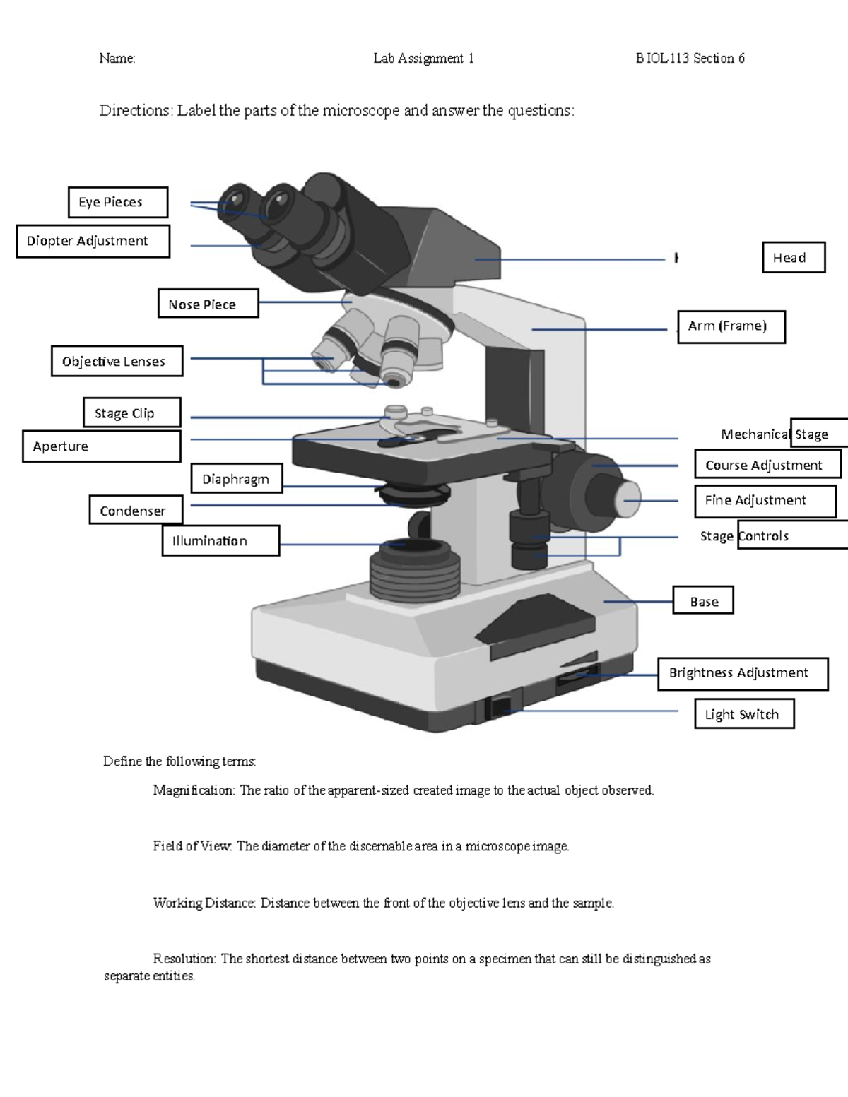
Microscopy Assignment - Name: Lab Assignment 1 BIOL113 ...
Light microscopes - Cell structure - Edexcel - BBC Bitesize The magnification of a lens is shown by a multiplication sign followed by the amount the lens magnifies. So a lens magnifying ten times would be ×10. The total magnification of a microscope is:...
Understand How to Use a Microscope Worksheet - EdPlace
The Eyes (Human Anatomy): Diagram, Optic Nerve, Iris, Cornea ... - WebMD Your eye is a slightly asymmetrical globe, about an inch in diameter. The front part (what you see in the mirror) includes: Iris: the colored part; Cornea: a clear dome over the iris; Pupil: the ...

Optical microscope, microscope, angle, simple, label png ...
Top 16 Techniques Used in Cell Biology (With Diagram) Then the bound labelled antigens are eluted from the column and can be quantitatively arranged through Liquid Scintillation counter. Antibodies are generally labelled with H 3,C 14 or I 131. When cells or tissues axe used, radiolabeled antibodies can be used with the help of autoradiography to localize the various components within the cell.

Draw a neat labelled diagram of a compound microscope class ...
Label the microscope — Science Learning Hub In this interactive, you can label the different parts of a microscope. Use this with the Microscope parts activity to help students identify and label the main parts of a microscope and then describe their functions. Drag and drop the text labels onto the microscope diagram.

Label the diagram of the microscope and explain the role of ...
Fluorescence - Wikipedia Fluorescence is the emission of light by a substance that has absorbed light or other electromagnetic radiation.It is a form of luminescence.In most cases, the emitted light has a longer wavelength, and therefore a lower photon energy, than the absorbed radiation.A perceptible example of fluorescence occurs when the absorbed radiation is in the ultraviolet region of the …

Diagram of a Compound Microscope
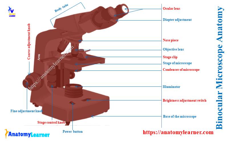
Binocular Microscope Anatomy - Parts and Functions with a Labeled Diagram The important non-optical parts of the light compound microscope are the body tube or head, arm or frame, fine adjustment, coarse adjustment, nose piece, stage, and base. Now, I will describe all these non-optical parts of the light compound microscope with the labeled diagrams. The body tube of the microscope
Instruments of Microscopy | Microbiology | | Course Hero
Parts of a Compound Microscope and Their Functions - NotesHippo Compound microscope mechanical parts (Microscope Diagram: 2) include base or foot, pillar, arm, inclination joint, stage, clips, diaphragm, body tube, nose piece, coarse adjustment knob and fine adjustment knob.. Base: It’s the horseshoe-shaped base structure of microscope.All of the other components of the compound microscope are supported by it. ...

microscope | Types, Parts, History, Diagram, & Facts | Britannica
PDF Label parts of the Microscope Label parts of the Microscope: . Created Date: 20150715115425Z
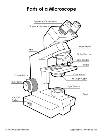
Microscope Diagram - Tim's Printables
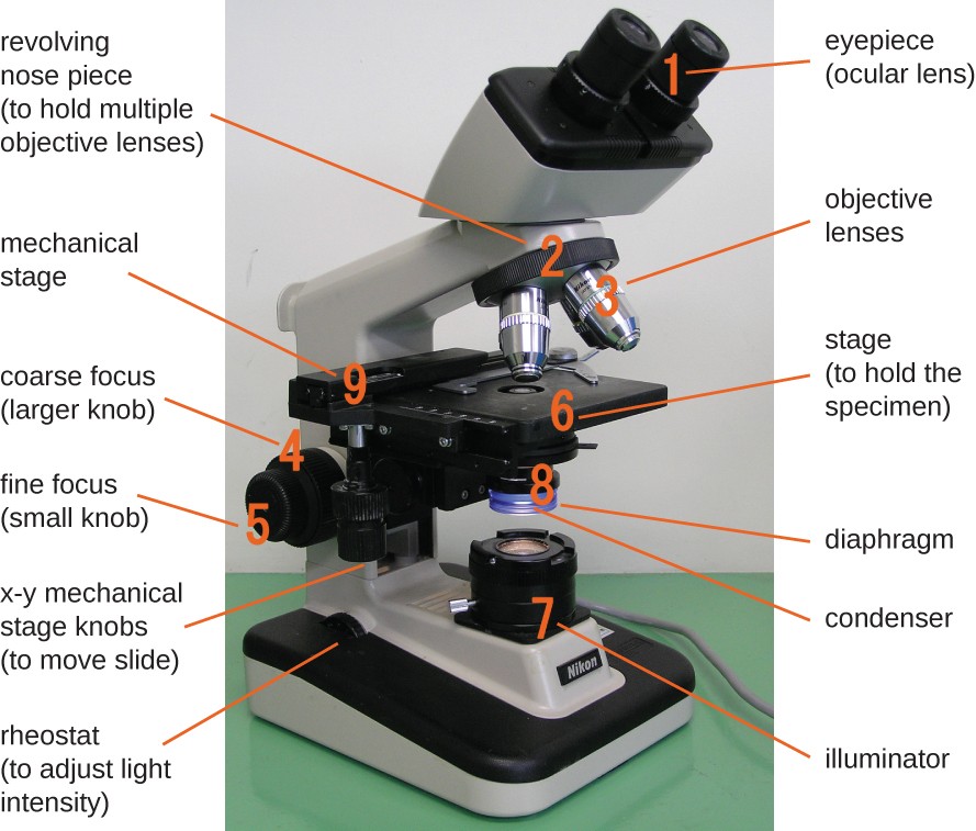
Parts of a microscope with functions and labeled diagram - Microbe Notes Q. List down the 18 parts of a Microscope. 1. Ocular Lens (Eye Piece) 2. Diopter Adjustment 3. Head 4. Nose Piece 5. Objective Lens 6. Arm (Carrying Handle) 7. Mechanical Stage 8. Stage Clip 9. Aperture 10. Diaphragm 11. Condenser 12. Coarse Adjustment 13. Fine Adjustment 14. Illuminator (Light Source) 15. Stage Controls 16. Base 17.
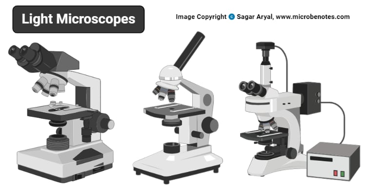
Light Microscope- Definition, Principle, Types, Parts ...
Parts of a Microscope Labeling Activity - Storyboard That Create a poster that labels the parts of a microscope and includes descriptions of what each part does. Click "Start Assignment". Use a landscape poster layout (large or small). Search for a diagram of a microscope. Using arrows and textables label each part of the microscope and describe its function.

BIOLOGY FROM 1 | EQUIPMENTS USED FOR OBSERVATION | Cours ...
Microscope labeled diagram - SlideShare 1. The Microscope Image courtesy of: Microscopehelp.com Basic rules to using the microscope 1. You should always carry a microscope with two hands, one on the arm and the other under the base. 2. You should always start on the lowest power objective lens and should always leave the microscope on the low power lens when you finish using it. 3.

Draw a labelled diagram of a plant cell as revealed by an electron microscope. | 9 | IMPROVEMEN...
Label Microscope Diagram - EnchantedLearning.com base - this supports the microscope. body tube - the tube that supports the eyepiece. coarse focus adjustment - a knob that makes large adjustments to the focus. diaphragm - an adjustable opening under the stage, allowing different amounts of light onto the stage. eyepiece - where you place your eye.

Microscopes: A Beginner's Guide
Label the microscope Diagram | Quizlet Diaphragm. Regulates the amount of light on the specimen. Light Source. Projects light upwards through the diaphragm, the specimen, and the lenses. Arm. supports the body tube. Stage. Supports the slide being viewed. Coarse Adjustment.
How to Use a Microscope
Light Microscope- Definition, Principle, Types, Parts, Labeled Diagram ... Figure: Labeled Diagram of a Light Microscope. Types of light microscopes (optical microscope) With the evolved field of Microbiology, the microscopes. used to view specimens are both simple and compound light microscopes, all using lenses. The difference is simple light microscopes use a single lens for magnification while compound lenses use ...

Draw a well labelled diagram of a microscope. - Brainly.in
Microscope Parts and Functions Most specimens are mounted on slides, flat rectangles of thin glass. The specimen is placed on the glass and a cover slip is placed over the specimen. This allows the slide to be easily inserted or removed from the microscope. It also allows the specimen to be labeled, transported, and stored without damage.
Lab :1
Compound Microscope Parts - Labeled Diagram and their Functions Labeled diagram of a compound microscope Major structural parts of a compound microscope There are three major structural parts of a compound microscope. The head includes the upper part of the microscope, which houses the most critical optical components, and the eyepiece tube of the microscope.

Simple Microscope - Diagram (Parts labelled), Principle ...
The Cell - ScienceQuiz.net Name the part of the cell labelled A in the diagram.? Cytoplasm? Membrane? Nucleus? Cell wall; The function of the cell membrane is? to control what enters and leaves the cell. ? to give the cell shape and support.? to control reproduction in the cell.? to control activities in the cell. Which one of the following is a nerve cell?? ? ? ? The process by which cells can make copies of …
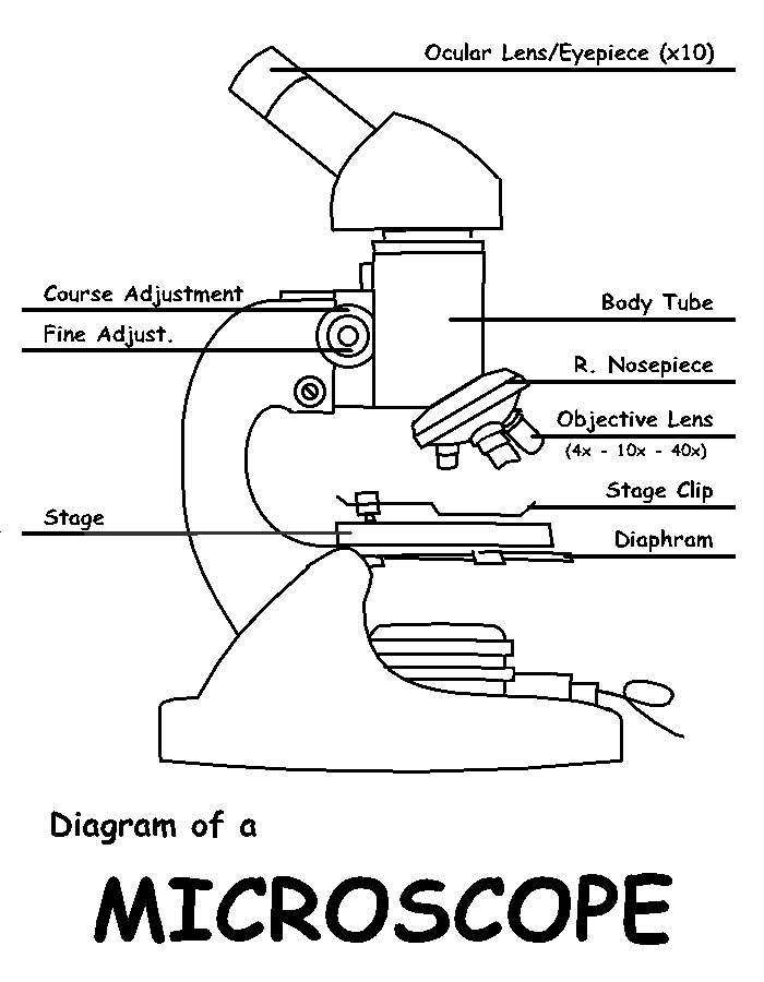
Diagram of a Microscope by ScienceDoodles on DeviantArt
Microscope Types (with labeled diagrams) and Functions Phase-contrast microscope labeled diagram Phase-contrast microscope functions: Its applications areas include In cases where the specimen is colorless and is very tiny In biology to conduct cellular level examination of microorganisms that can't be visualized using the bright field microscopy Interference Microscope

This is a common compound microscope. Label its parts from A ...
Investigating cells with a light microscope - BBC Bitesize Record the microscope images using labelled diagrams or produce digital images. When first examining cells or tissues with low power, draw an image at this stage, even if going on to examine the ...
Binocular Microscope Anatomy - Parts and Functions with a ...
Microscope Parts, Function, & Labeled Diagram - slidingmotion Microscope parts labeled diagram gives us all the information about its parts and their position in the microscope. Microscope Parts Labeled Diagram The principle of the Microscope gives you an exact reason to use it. It works on the 3 principles. Magnification Resolving Power Numerical Aperture. Parts of Microscope Head Base Arm Eyepiece Lens

File:Microscope diagram.png - Wikimedia Commons

File:Labelledmicroscope.gif - Wikimedia Commons

Label Microscope Diagram - EnchantedLearning.com

Microscope World Blog: High School Microscope Features
Microscope drawing - Teaching resources
Draw a labelled ray diagram of a compound microscope and ...

Microscope Labeling Diagram | Quizlet

Compound Microscope Parts – Labeled Diagram and their ...

Microscope - Label - Part 2 Diagram | Quizlet

Compound Microscope: Definition, Diagram, Parts, Uses ...

Parts of a microscope with functions and labeled diagram

Microscope Labeling
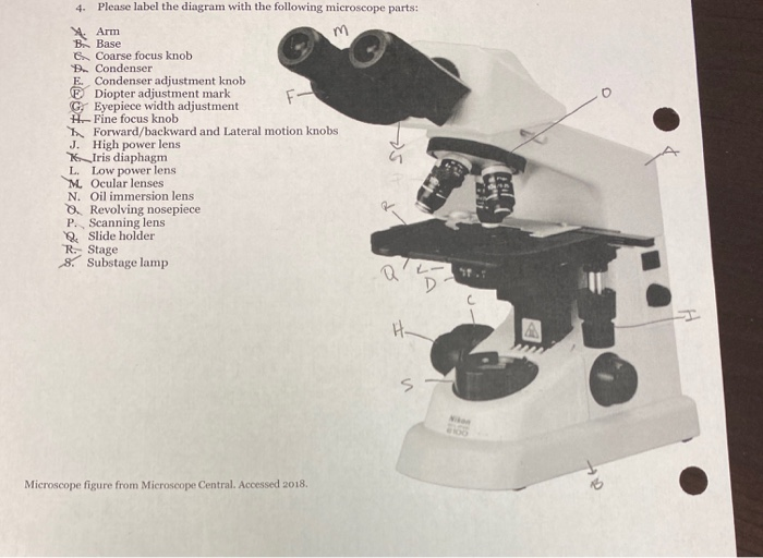
Solved 4. Please label the diagram with the following | Chegg.com
1.2: Microscopes - Biology LibreTexts

Label the microscope — Science Learning Hub
Simple Microscope - Parts, Functions, Diagram and Labelling ...

The Microscope
Microscopes - SCIENTIST CINDY
Untitled Document

Microscope Parts and Functions

Microscope Maintenance Tips | Science supplies, Multi step ...
Using Microscopes - Bio111 Lab
Post a Comment for "45 labelled diagram of microscope"