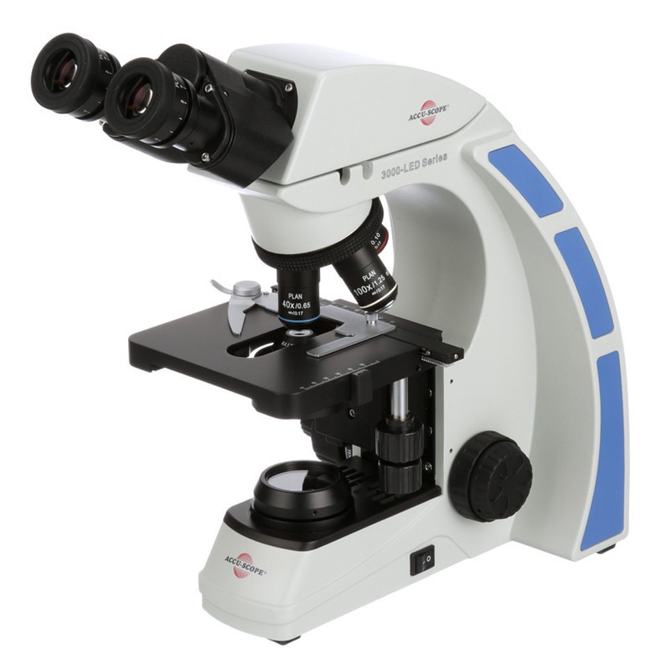
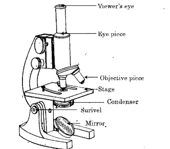
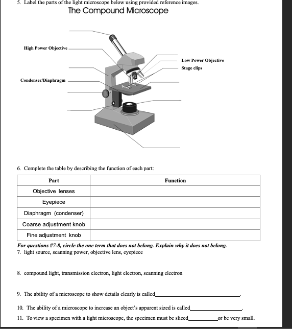
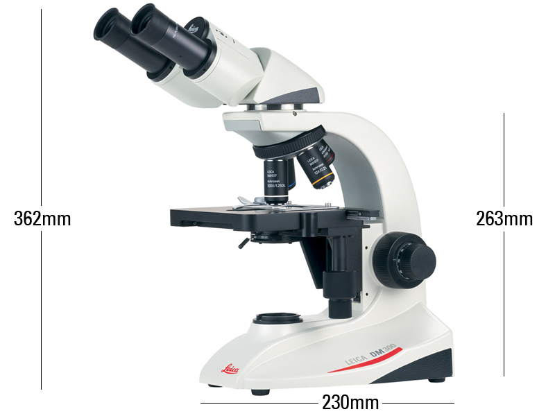
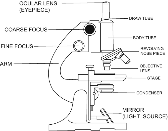
41 compound microscope labeled
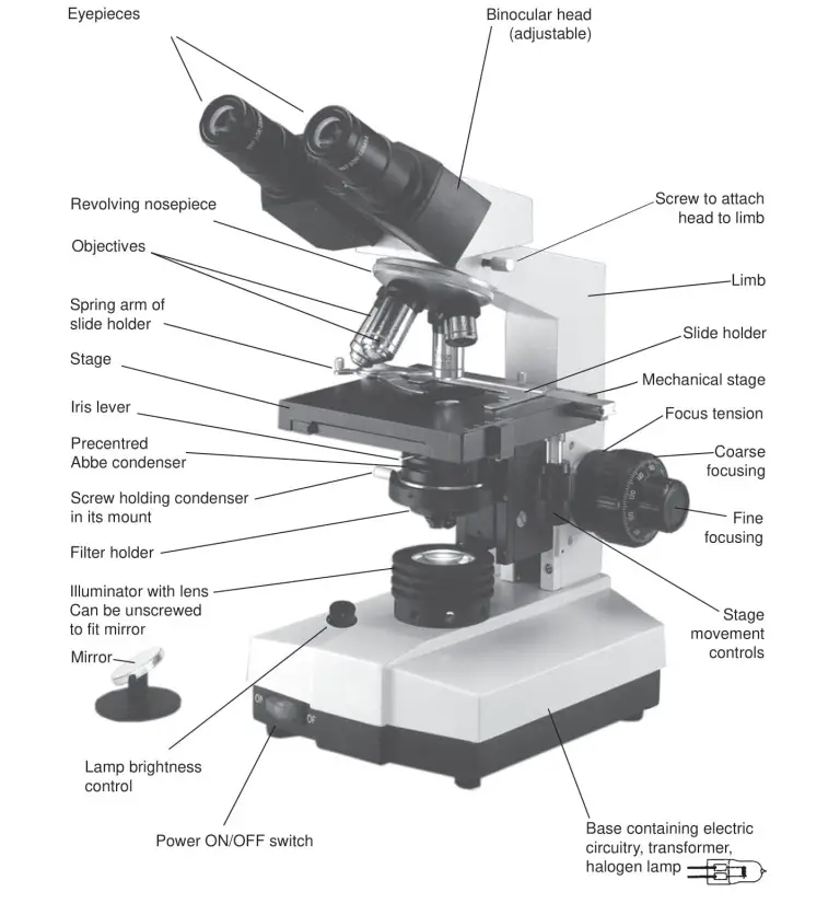
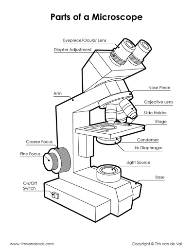
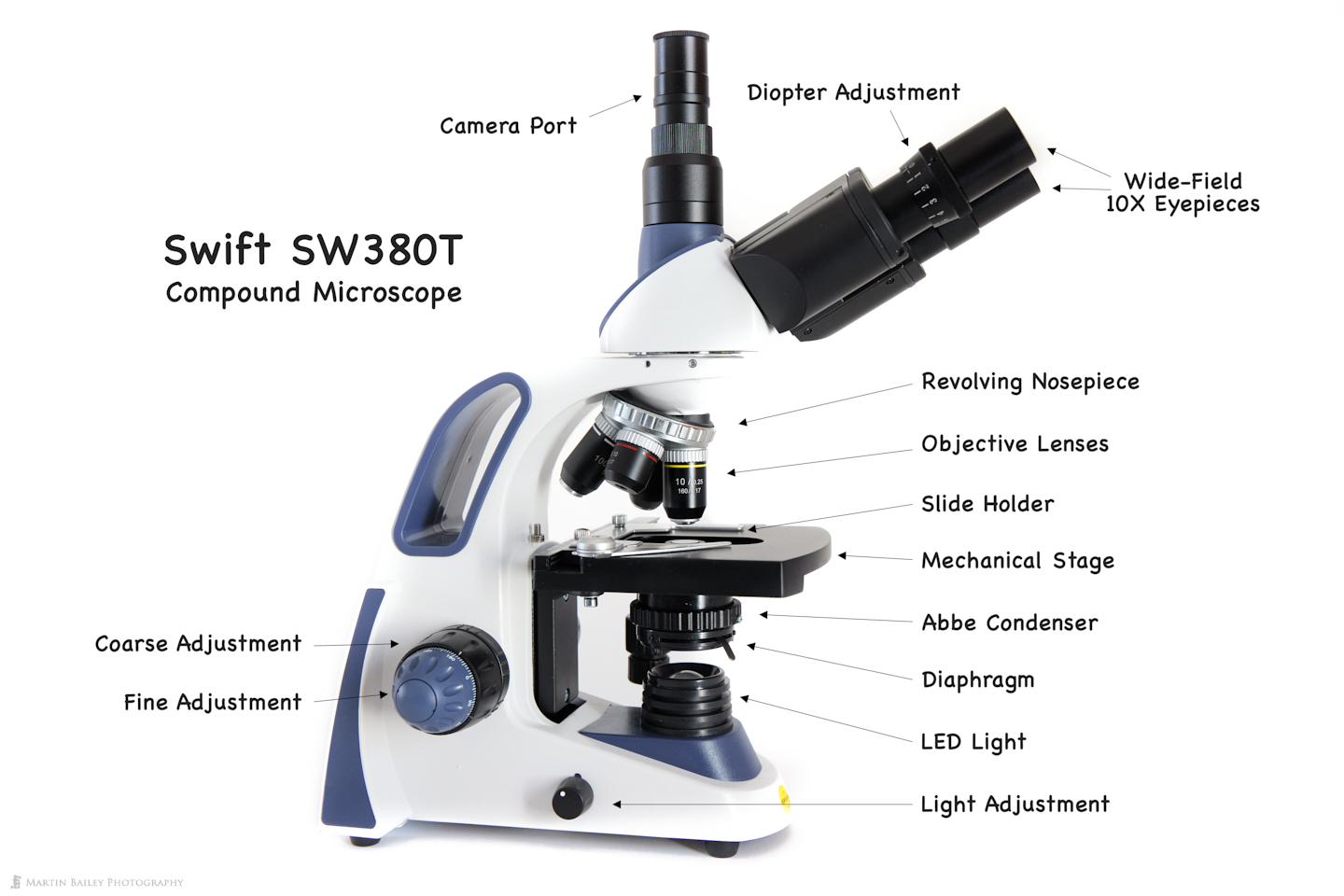
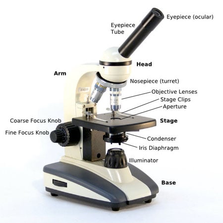
Parts of a microscope with functions and labeled diagram -... Sep 17, 2022 · For monocular microscopes, they are none flexible. Objective lenses – These are the major lenses used for specimen visualization. They have a magnification power of 40x-100X. There are about 1- 4 objective lenses placed on one microscope, in that some are rare facing and others face forward. Each lens has its own magnification power. Compound Microscope – Diagram (Parts labelled), Principle and... Oct 10, 2022 · Why is it called compound microscope? Because it has multiple lenses that work in conjunction to magnify a specimen Q 4. What are the 13 parts of a microscope? 1. Eyepiece 2. Eyepiece Tube 3. Objective Lens 4. Stage 5. Stage Clips 6. Nosepiece 7. Fine and Coarse Focus knobs 8. Illuminator 9. Aperture 10. Iris Diaphragm 11. Condenser 12.
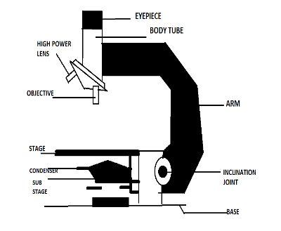
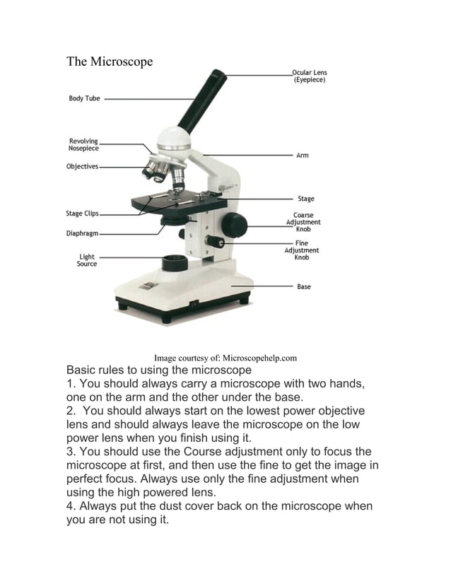
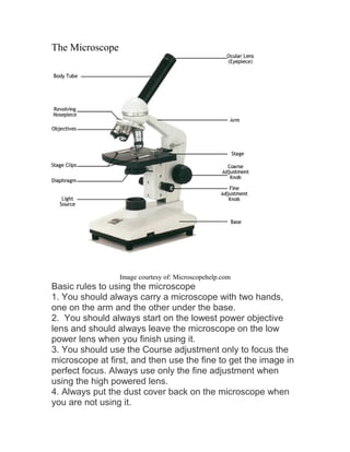
Compound Microscope Labeled Diagram | Quizlet Compound Microscope Labeled + − Flashcards Learn Test Match Created by meganplocher734 Terms in this set (14) Eyepiece/Ocular lens Contains the ocular lens Body tube A hollow cylinder that holds the eyepiece. Arm Part that supports the microscope. Stage Supports the slide or specimen Coarse adjustment Knob

Compound microscope labeled
Labelled Diagram of Compound Microscope The below mentioned article provides a labelled diagram of compound microscope. Part # 1. The Stand: The stand is made up of a heavy foot which carries a curved inclinable limb or arm bearing the body tube. The foot is generally horse shoe-shaped structure (Fig. 2) which rests on table top or any other surface on which the microscope in kept. Label the microscope — Science Learning Hub Jun 8, 2018 · Label the microscope Interactive Add to collection Use this interactive to identify and label the main parts of a microscope. Drag and drop the text labels onto the microscope diagram. eye piece lens diaphragm or iris coarse focus adjustment stage base fine focus adjustment light source high-power objective Download Exercise Tweet Parts of a Compound Microscope - Labeled (with diagrams) Mar 6, 2020 · Parts of a Compound Microscope - Labeled (with diagrams) A compound microscope is known as a high-power microscope that enables you to achieve a high level of magnification. Smaller specimens can be thoroughly viewed using a compound microscope. Let us take a look at the different parts of a compound microscope and understand each key component. Image 1: The figure above is the standard image of a compound microscope.
Compound microscope labeled. Compound Microscope: Definition, Diagram, Parts, Uses, Working... A compound microscope is defined as. A microscope with a high resolution and uses two sets of lenses providing a 2-dimensional image of the sample. The term compound refers to the usage of more than one lens in the microscope. Also, the compound microscope is one of the types of optical microscopes. The other type of optical microscope is a simple microscope. Parts of a Compound Microscope - Labeled (with diagrams) Mar 6, 2020 · Parts of a Compound Microscope - Labeled (with diagrams) A compound microscope is known as a high-power microscope that enables you to achieve a high level of magnification. Smaller specimens can be thoroughly viewed using a compound microscope. Let us take a look at the different parts of a compound microscope and understand each key component. Image 1: The figure above is the standard image of a compound microscope. Label the microscope — Science Learning Hub Jun 8, 2018 · Label the microscope Interactive Add to collection Use this interactive to identify and label the main parts of a microscope. Drag and drop the text labels onto the microscope diagram. eye piece lens diaphragm or iris coarse focus adjustment stage base fine focus adjustment light source high-power objective Download Exercise Tweet Labelled Diagram of Compound Microscope The below mentioned article provides a labelled diagram of compound microscope. Part # 1. The Stand: The stand is made up of a heavy foot which carries a curved inclinable limb or arm bearing the body tube. The foot is generally horse shoe-shaped structure (Fig. 2) which rests on table top or any other surface on which the microscope in kept.











Post a Comment for "41 compound microscope labeled"