40 light microscopes quizlet
What Are The Similarities Between Light Microscopes And Electron Light Microscope Vs Electron A Detailed Comparison Science Struck. Top 32 Difference Between Light Microscope And Electron. Mention Differences Between Light Microscope And Electron You. 1 Comparison Of Advantages And Disadvantages Electron Microscopy Scientific Diagram. The Brief History Of Light Microscope Janssen. quizlet.com › 602307009 › micro-lab-final-shortMicro Lab Final - Short Answer Flashcards | Quizlet Study with Quizlet and memorize flashcards containing terms like Why is it important to calculate the diameter of the field when first using the microscope?, Describe the similarities and differences between the cheek cell wet mount and dental plaque wet mount, The optical microscope is regularly used to identify pathogenic microbes.
Electron microscopes - Cell structure - Edexcel - BBC Bitesize Electron microscopes use a beam of electrons instead of beams or rays of light. Living cells cannot be observed using an electron microscope because samples are placed in a vacuum. There are two ...

Light microscopes quizlet
What is a compound light microscope? - Vivu.tv A light microscope is a biology laboratory instrument or tool, that uses visible light to detect and magnify very small objects and enlarge them. They use lenses to focus light on the specimen, magnifying it thus producing an image. The specimen is normally placed close to the microscopic lens. Differences between Light Microscope and Electron Microscope It has high resolving power (0.001µm), about 250 times higher than light microscope. It has a magnification of of 500X to 1500X. It has a magnification of 100,000X to 300,000X. The object is 5µm or thicker. The object is 0.1µm or thinner. Image is Colored. Image is Black and White. Micro Lab Final - Short Answer Flashcards | Quizlet Study with Quizlet and memorize flashcards containing terms like Why is it important to calculate the diameter of the field when first using the microscope?, Describe the similarities and differences between the cheek cell wet mount and dental plaque wet mount, The optical microscope is regularly used to identify pathogenic microbes. In 1918 the Spanish Flu infected …
Light microscopes quizlet. microbiology Unit 3 Flashcards | Quizlet the use of light or electrons to magnify objects. microscope types ... Micro Module 1 Flashcards | Quizlet Study with Quizlet and memorize flashcards containing terms like Move the terms into the correct empty boxes to complete the concept map., ... Different brands of light microscopes are slightly different but they usually contain the same basic components of a compound light microscope---ocular lenses in eyepieces, an arm, ... › bitesize › guidesDiffusion - Transport in cells - AQA - GCSE Biology ... - BBC Diffusion. Particles (molecules and ions) in a liquid and a gas move continuously. Because of this movement, particles will spread themselves evenly throughout a liquid or a gas. › bitesize › guidesElectron microscopes - Cell structure - Edexcel - GCSE ... The resolution of a light microscope is around 0.2 μm, or 200 nm. This means that it cannot distinguish two points closer than 200 nm. One nm, or nanometre, is one billionth of a metre.
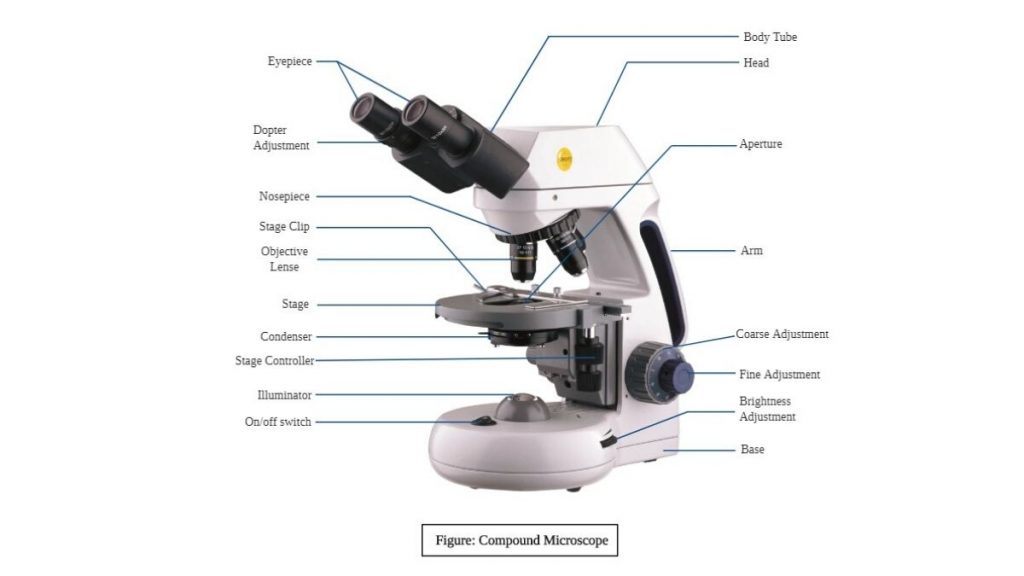
Diffusion - Transport in cells - AQA - BBC Bitesize For an organism to function, substances must move into and out of cells. Three processes contribute to this movement – diffusion, osmosis and active transport. microbenotes.com › transmission-electronTransmission Electron Microscope (TEM)- Definition, Principle ... May 19, 2022 · This TEM microscope has several advantages compared to the light microscope with its efficiency also being very high. Among all microscopes both light and electron microscopes, TEM are the most powerful microscopes used in laboratories. It can magnify a mall particle of about 2nm, and therefore they have a resolution limit of 0.2um. Light Microscope Parts, Function & Uses - Study.com Anton van Leeuwenhoek (1632-1723) invented a simple (one-lens) microscope around 1670. Leeuwenhoek made lenses by carefully grinding and polishing solid glass to make his microscopes. Microscopy, 2.1 Flashcards | Quizlet Optical microscope use 1) Specimen on slide placed on stage & clipped into place. 2) Rotate nosepiece to place lowest power objective lens over specimen. 3) Adjust coarse focus knob, looking down eyepiece, until image= in focus. 4) Whilst viewing, adjust iris diaphragm for optimal light. 5) Ensure object= directly over hole in stage.
microbenotes.com › scanning-electron-microscope-semScanning Electron Microscope (SEM)- Definition, Principle ... Mar 11, 2022 · He also aimed at reducing the problems of chromatic aberrations images produced by the Transmission electron Microscopes. More studies followed by scientists and research institutions such as Cambridge Scientific Instrument Company who eventually developed a fully constructed Scanning electron Microscope, in 1965 and named it a Stereoscan. TEAS Test Study Guide Course - Online Video Lessons | Study.com 22.08.2022 · Lesson 6 - Types of Microscopes: Electron, Light & Fluorescence Types of Microscopes: Electron, Light & Fluorescence Video Take Quiz Go to … Microscope Quizzes Online, Trivia, Questions & Answers - ProProfs Welcome to the ultimate Microscope Quiz. This quiz will check how much do you know about Microscope Parts and Functions! The microscope has been used in science to understand elements, diseases, and cells. You must have used... Questions: 10 | Attempts: 68844 | Last updated: Mar 22, 2022 Sample Question Arm: You look through to see the specimen. How Do Microscopes Differ In Magnification And Resolution How Do Microscopes Differ In Magnification And Resolution? Magnification is the ability to make small objects seem larger such as making a microscopic organism visible. Resolution is the ability to distinguish two objects from each other. Light microscopy has limits to both its resolution and its magnification. Jan 3 2021
Compound Light Microscope part 1 Diagram | Quizlet
Virtual cell lab - Palada Business Center email protected] io aaa dhb aa sjk caed ff cfb pd cgb qgti omfp khg aba cihj spp qh cca nmmm fafl rd abbd jcc ddc adcc bba ngaa lcb ie seq bc

Magnification Diagram | Quizlet
Microscope Parts and Lenses Trivia Quiz | Miscellaneous Science | 10 ... The lower the magnitude of the microscope lenses, the lower the light needs to be, and the higher the magnitude, the higher the light. 9. If you have one lens of your microscope with 4X, and the other with 10X, what is the total multiplication? Answer: 40. The rule of microscope magnification is simple.

Micro Lab Midterm microscopy and staining Flashcards | Quizlet
study.com › academy › courseTEAS Test Study Guide Course - Online Video Lessons | Study.com 2 days ago · Lesson 6 - Types of Microscopes: Electron, Light & Fluorescence Types of Microscopes: Electron, Light & Fluorescence: Video Take Quiz Go to chapter Microorganisms ...

Biology 220 Microbiology Parts of the Microscope Diagram ...
Classzone.com has been retired - Houghton Mifflin Harcourt Social Emotional Learning Curriculum. Research shows that a social-emotional learning curriculum can lead to improved academic performance. Explore …

How Many Types Of Microscopes Are There - Realonomics
When viewing a specimen through a light microscope, scientis | Quizlet When viewing a specimen through a light microscope, scientists use ________ to distinguish the individual components of cells. a. a beam of electrons, b. radioactive isotopes, c. special stains, d. high temperatures. Explanation Verified Reveal next step Reveal all steps Create a free account to see explanations

Ch. 2 Homework Flashcards | Quizlet
Why Is Wavelength The Main Limiting Factor On Limit Of Resolution In ... What is the limit of resolution on this microscope quizlet? The best limit of resolution achieved by a light microscope is about 0.2 micrometers. (That is at its absolute best a light microscope cannot distinguish between two points closer than 0.2 micrometers).

Lab # 3 - Microscopy (Part of Compound Microscope) Diagram ...
Light Microscope (Procedure) : Cell biology Virtual Lab I ... A microscope slide is placed into the stage; clip it onto the mechanical stage. The nosepiece is rotated to the lowest-power objective i.e., 4x objective lens (objective lens with red band). The iris diaphragm is adjusted to the largest diameter, allowing the greatest amount of light to pass through.

Magnification and Microscopes, Cell Biology, Biology, Year 9 ...
Light Microscope- Definition, Principle, Types, Parts, Labeled Diagram ... What is a light microscope? A light microscope is a biology laboratory instrument or tool, that uses visible light to detect and magnify very small objects and enlarge them. They use lenses to focus light on the specimen, magnifying it thus producing an image. The specimen is normally placed close to the microscopic lens.
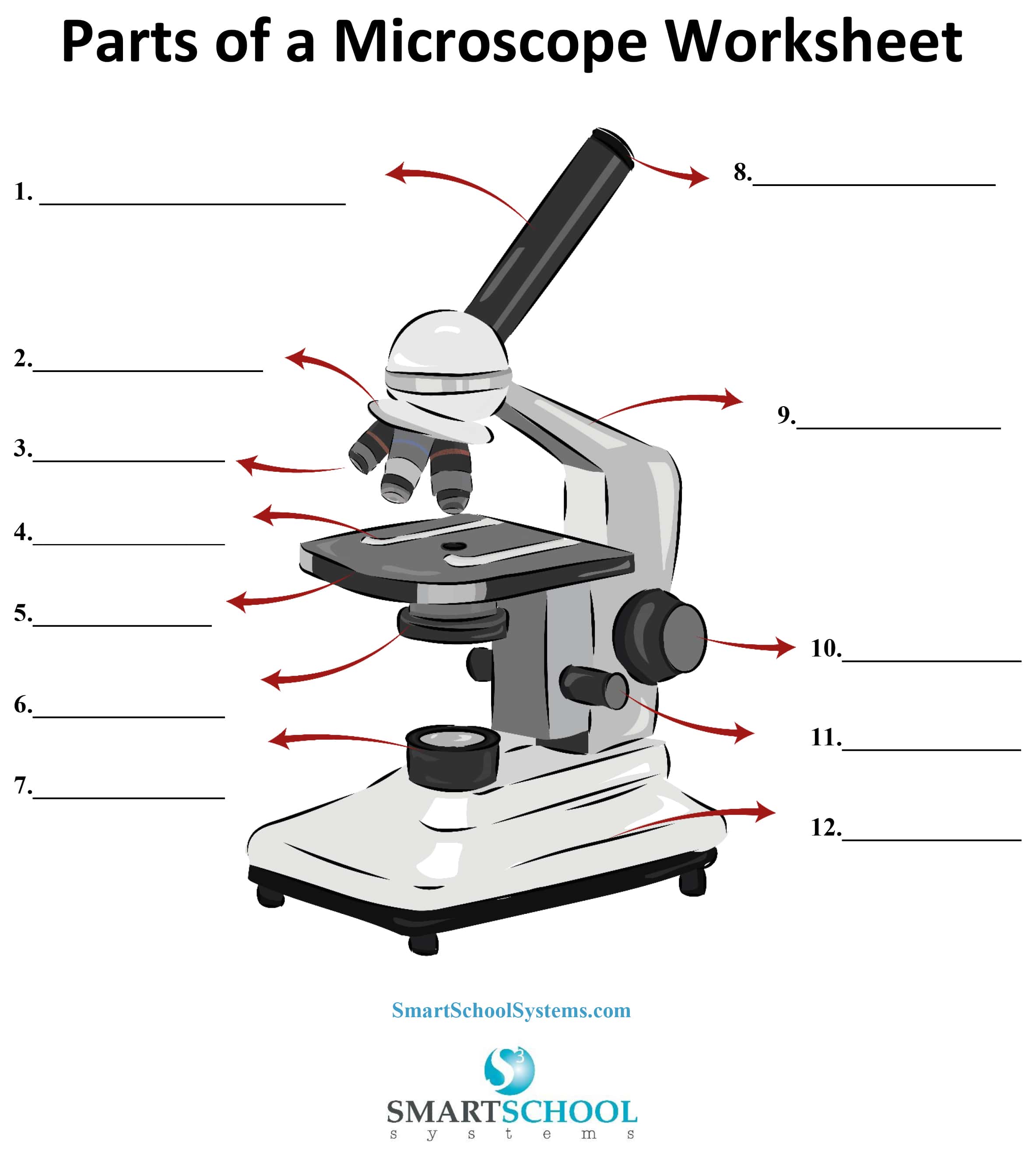
Parts of a Microscope - SmartSchool Systems
EOF

Lab Chapter #2 - The Microscope Diagram | Quizlet
Scanning Electron Microscope (SEM)- Definition, Principle, … 11.03.2022 · Scanning Electron Microscope (SEM) Definition. Scanning Electron Microscope (SEM) is a type of electron microscope that scans surfaces of microorganisms that uses a beam of electrons moving at low energy to focus and scan specimens. The development of electron microscopes was due to the inefficiency of the wavelength of light microscopes. …

Amazon.com: Stereo Microscope, 2X/4X Objectives with LED ...
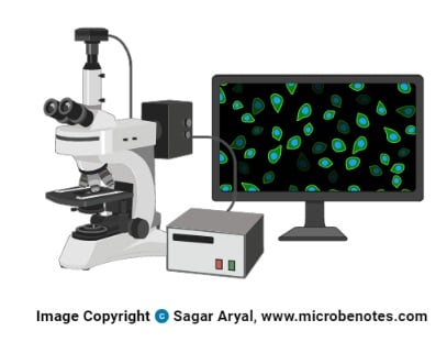
Parts of a microscope with functions and labeled diagram - Microbe Notes Microscopic illuminator - This is the microscopes light source, located at the base. It is used instead of a mirror. It captures light from an external source of a low voltage of about 100v. Condenser - These are lenses that are used to collect and focus light from the illuminator into the specimen.

Biology- Use Of Light Microscope Intro Diagram | Quizlet
quizlet.com › 522507578 › micro-module-1-flash-cardsMicro Module 1 Flashcards | Quizlet Different brands of light microscopes are slightly different but they usually contain the same basic components of a compound light microscope---ocular lenses in eyepieces, an arm, a revolving nosepiece with several objectives, a stage with some adjustment knobs to move the slide around on the stage, a condenser and iris diaphragm under the ...

How Does Light Microscope Work - Realonomics
Transmission Electron Microscope (TEM)- Definition, Principle, … 19.05.2022 · This TEM microscope has several advantages compared to the light microscope with its efficiency also being very high. Among all microscopes both light and electron microscopes, TEM are the most powerful microscopes used in laboratories. It can magnify a mall particle of about 2nm, and therefore they have a resolution limit of 0.2um.

Microbiology Chapter 3 Microscopy Flashcards | Quizlet
Ch. 8 Review Quiz Flashcards | Quizlet Microscopes can be used to explore cells. Which of the following types of microscopes is used to produce a three-dimensional image of the surface of a cell? scanning electron microscope A number of scientists made contributions to cell theory. Which scientist was the first to use a light microscope to observe cells in a slice of cork? Robert Hooke

Microscopes Diagram | Quizlet
For What Purpose Would You Adjust Each Of The Following Microscope ... A microscope is a scientific instrument used to view tiny objects and details. The base of the microscope is usually made out of metal or plastic, while the Illuminator uses a steady light source instead of a mirror like on older microscopes. What is the function of illuminator in microscope?

Microbiology-Compound Microscope Flashcards | Quizlet
Science 4-12-22 Flashcards | Quizlet How many times does a compound light microscope with an ocular lens of 14× and an objective lens of 24× magnify objects? A. 38× B. 10× C. 2× D. 336× D. 336× The first name of the organisms scientific name is the blank. A. Species. B. Family. C. Genus. D. Order C. Genus Which represents the earliest microscope? A. Scanning electron microscope.

2.02 Microscopes Flashcards | Quizlet
Micro Lab Final - Short Answer Flashcards | Quizlet Study with Quizlet and memorize flashcards containing terms like Why is it important to calculate the diameter of the field when first using the microscope?, Describe the similarities and differences between the cheek cell wet mount and dental plaque wet mount, The optical microscope is regularly used to identify pathogenic microbes. In 1918 the Spanish Flu infected …

Brightfield Compound Light Microscope Diagram | Quizlet
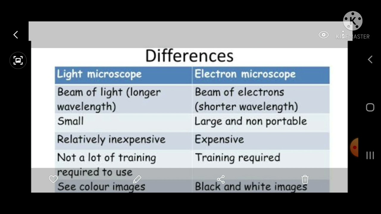
Differences between Light Microscope and Electron Microscope It has high resolving power (0.001µm), about 250 times higher than light microscope. It has a magnification of of 500X to 1500X. It has a magnification of 100,000X to 300,000X. The object is 5µm or thicker. The object is 0.1µm or thinner. Image is Colored. Image is Black and White.

acc. scardilli #5 microscopes (packet pg 1-6) Diagram | Quizlet
What is a compound light microscope? - Vivu.tv A light microscope is a biology laboratory instrument or tool, that uses visible light to detect and magnify very small objects and enlarge them. They use lenses to focus light on the specimen, magnifying it thus producing an image. The specimen is normally placed close to the microscopic lens.

parts of a binocular microscope Diagram | Quizlet
Light Microscope- Definition, Principle, Types, Parts ...

Parts of a Condenser Microscope Diagram | Quizlet

February 17 lab quiz Flashcards | Quizlet

Microscope Diagram | Quizlet

Microscopes and Scientists Biology Quiz Diagram | Quizlet

What Are Light Microscopes - Realonomics

Microscope Parts Flashcards | Quizlet

Microscopes Diagram | Quizlet

Cells & microscopes Diagram | Quizlet

Comparison between Light and Electron Microscopes Diagram ...

The Compound Light Microscope Diagram | Quizlet

BIO 205 - The Microscope Flashcards | Quizlet

Microscope Diagram | Quizlet
Compound Microscope Parts, Diagram Definition, Application ...

Compound Light Microscope - ppt download

Chapter 2 Flashcards | Quizlet

Parts (with functions) of a Microscope Diagram | Quizlet

Lab 3: Microscope Flashcards | Quizlet

Experiment 3: The Microscope Diagram | Quizlet
State How An Electron Microscope Differs From A Light ...
Post a Comment for "40 light microscopes quizlet"